
Page 33 - Summer 2010
P. 33
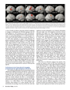
  Fig. 4. Significant functional magnetic resonance imaging (fMRI) activations in a single patient before and after therapy. a) Activations before therapy. b) Activations after therapy. c) A direct comparison between after-therapy and before-therapy activations. The top panels show left and right brain activation during overt speaking contrasted with a silent control. The bottom panels show brain activation during overt speaking contrasted with the control task of vowel production. All contrasts are superimposed on a spatially standardized normal brain template and use a threshold at a level of p < 0.05 (statistically corrected for family-wise error). The color codes represent different magnitudes of activation: stronger activations are indicated in yellow. Reprinted with permission from Schlaug et al. (2010).
a series of words or phrases using slow, pitched vocalization or singing in combination with rhythmic tapping of the left hand (Albert et al., 1973; Norton et al., 2009). The effective- ness of MIT has been demonstrated by behavioral improve- ments in naming (Bonakdarpour et al., 2000; Sparks et al., 1974) and in articulation and phrase production (Wilson et al., 2006) after treatment. A recent study involving two patients examined the benefit of MIT on transfer of language skills to untrained contexts (Schlaug et al., 2008). In that study, which involved 75 daily sessions of 90 minutes, the extent of this improvement was far greater for the patient who underwent MIT compared to the one who underwent the control therapy. Furthermore, neuroimaging of this patient showed that MIT results in increased activation in a right-hemisphere network involving the premotor, inferior frontal, and temporal lobes (Schlaug et al., 2008), as well as increased fiber number and volume of the arcuate fasciculus in the right hemisphere (Schlaug et al., 2009). Critically, the patient treated with a non-intonation-based speech therapy showed smaller right hemisphere changes and more left hemisphere changes. These findings demonstrate that inten- sive training involving through singing, applied over a longer period of time in chronic stroke patients, can induce func- tional and structural brain changes. These changes are relat- ed to speech output improvements in patients suffering from aphasia.
Facilitating recovery from aphasia by engaging predominantly the right hemisphere of the brain
The traditional explanation for the dissociation between speaking and singing in patients with aphasia is the presence of two routes for word articulation: one for spoken words through the brain’s left hemisphere, and a separate route for sung words that uses either the right or both hemispheres. However, research indicates that there is a bi-hemispheric role in the execution and sensorimotor control of vocal pro- duction for both speaking and singing (Guenther et al., 1998; Jeffries et al., 2003; Brown et al., 2004; Bohland and Guenther, 2006; Ozdemir et al., 2006), typically with a left- lateralization for speaking. It has been shown that tasks that
emphasize spectral information over temporal information elicit more right- than left-hemispheric activation (Zatorre and Belin, 2001; Meyer et al., 2002). Similarly, patients with right-hemisphere lesions have greater difficulty with global processing (e.g., melody and contour processing) compared to those with left-hemisphere lesions (Peretz, 1990; Schuppert et al., 2000). Thus, it is possible that the melodic element of MIT engages the right hemisphere, particularly the right temporal lobe, more so than therapies that do not make use of pitch or melody. To date, only a handful of brain imaging studies has shed light on the possible neural mecha- nisms underlying language recovery following MIT. One interpretation is that MIT engages the expressive language areas in the right hemisphere. Alternatively, MIT could exert its effect either by unmasking existing music/language con- nections in both hemispheres, or by engaging preserved lan- guage-capable regions in either or both hemispheres. Since MIT incorporates both the melodic and rhythmic aspects of music (Albert et al., 1973; Sparks et al., 1974; Sparks and Holland, 1976; Helm-Estabrooks, 1989; Cohen and Masse, 1993; Boucher et al., 2001; Norton et al., 2009), it may be unique in its potential to engage not only the right, but both hemispheres.
Data from our laboratory indicate that patients who undergo an intensive course of MIT show functional and structural brain changes in their right hemispheres (Schlaug et al., 2008; Schlaug et al., 2009). Figure 4 shows significant fMRI activations in a single patient before and after therapy (Fig. 4a: before therapy; Fig. 4b: after therapy). The top pan- els show left and right brain activation during overt speaking contrasted with a silent control. The bottom panels show brain activation during overt speaking contrasted with the control task of vowel production. All contrasts are superim- posed on a spatially standardized normal brain template and thresholded at a level of p < 0.05 (statistically corrected for family-wise error.) Furthermore, a direct voxel-by-voxel comparison of the two acquisitions is shown in Fig. 4c. The color codes represent different magnitudes of activation: stronger activations are indicated in yellow. As shown in this figure, the most significant activations in these patients dur-
32 Acoustics Today, July 2010
