
Page 44 - Volume 12, Issue 2 - Spring 2012
P. 44
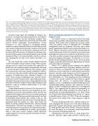
  Fig. 1. (A) Average normalized frequency tuning of excitation and inhibition from voltage clamp recordings for pre-nucleus basalis pairing. Synaptic currents were normal- ized to the amplitude of the largest EPSC (excitatory postsynaptic conductance) and IPSC (inhibitory) across frequencies. The center frequency (0 octaves) was set to the best frequency (BF) of excitation. Filled symbols, excitation; open symbols, inhibition. Error bars=sem. (B) Example excitatory frequency tuning curves based on measurements of synaptic conductance for an AI cell. Excitatory conductance at the paired frequency (2 kHz) increased from 0.9±0.2 nS to 1.6±0.1 nS (77.8%, p<0.006). Mean tuning curves before (dashed) and 10 min after (solid) plasticity induction. Down arrow=frequency of the paired tone; up arrow=pre-induction BF. (C) Example inhibitory fre- quency tuning curves based on measurements of synaptic conductance for same cell as shown in (B). Inhibitory conductance at the paired frequency (2 kHz) decreased from 1.4±0.3 nS to 0.6±0.2 nS (–57.1%, p<0.04).
Recently, using whole-cell recordings of synaptic con- ductances, we found that long-lasting positive and negative changes to auditory cortical excitatory synapses were induced, if acoustic stimuli were paired with activation of the nucleus basalis cholinergic neuromodulatory system (Froemke et al., 2007). Pairing of a sub-optimal stimulus inside the receptive field with release of acetylcholine for only a few minutes enhanced the excitatory response to the paired frequency (Fig. 1B), but decreased the excitatory activity at the former BF of the neurons. In addition, the inhibitory con- ductance of the neurons at the paired frequency was reduced temporarily (Fig. 1C), but recovered after several tens of min- utes matching the new BF of the neuron (Froemke et al., 2007).
We also found that nucleus basalis-enabled plasticity could be extended to shape intensity tuning. When synaptic tuning curves for sound level intensity were examined, we found that tone-evoked excitation monotonically increased as sound level grew louder. However, after pairing a quiet, low-intensity tone with nucleus basalis stimulation, respons- es to soft tones that were initially weak became stronger, and responses to louder tones were depressed (Carcea et al., 2012). These synaptic modifications were precisely orches- trated across entire receptive fields, conserving mean excita- tion while reducing overall variance, which implied that each parameter of cortical synaptic receptive fields (frequency and intensity) could be modified independently of the other (Carcea et al., 2012).
Computational analysis indicated that decreased vari- ability should increase detection and recognition of near- threshold or previously imperceptible stimuli and this was confirmed psychophysically with nucleus basalis pairing in behaving animals. Pairing in anesthetized animals can lead to behavioral improvements after animals woke up. The effects of pairing lasted only a few hours unless pairing was performed daily for several (6+) days, after which the effects of pairing endured. Furthermore, pharmacological manipulations indicated that changes to auditory cortex were both necessary and sufficient for behavioral enhance- ment. Thus, direct modification of specific cortical inputs leads to wide-scale synaptic changes, which collectively support improved sensory perception and enhanced behav- ioral performance.
Micro-organization and plasticity of the primary auditory cortex
The auditory cortex is a laminated structure that adap- tively processes auditory information from the external envi- ronment. Prior studies have revealed that on large spatial scales the auditory cortex in mammals has a tonotopic arrangement based on frequency selectivity and a more patchy organization of other response properties (Read et al., 2002). However, the precise nature of the transformation of sensory information at the level of auditory cortical networks and single cortical neurons is unknown. In the last few years, it has been possible to monitor large populations of neurons using new imaging technologies. In particular, in vivo two- photon calcium imaging techniques can be used to measure response properties and functional organization of cortical areas with single cell resolution.
When in vivo, 2-photon calcium techniques were applied to the supragranular layers of primary auditory cortex (A1) neurons in mouse, we found that the large scale tonotopic architecture of A1 breaks down at smaller scales (around 300 microns), and that nearby frequency and level tuning prop- erties were quite heterogeneous (Fig. 2; Bandyopadhyay et al., 2010, Rothschild et al., 2010). Imaging with dyes of differing sensitivity for calcium enabled the observation of signals arising from two sources: (1) suprathreshold spiking responses, or (2) a combination of subthreshold inputs and suprathreshold spiking responses. The comparison of these signals revealed a pronounced difference in heterogeneity (Fig. 2). This suggested that the observed heterogeneity on small spatial scales is likely created by the diverse inputs to supragranular neurons (Bandyopadhyay et al., 2010), which was recently observed by imaging sound driven Ca2+ signals in dendritic spines (Chen et al., 2011). Thus each supragran- ular neuron may sample from a large frequency range of inputs.
There are multiple possible intra- and inter-laminar net- work topologies that can give rise to the observed hetero- geneity. For example, intra-laminar connectivity within layer 2/3 could provide input from “distant” frequency channels. To reveal the inputs to A1 networks, current experiments have focused on stimulating single cells.
Supragranular cells have access to a large range of fre- quencies, and these inputs might provide a substrate for a
Auditory Cortical Function 43
