
Page 24 - 2017Spring
P. 24
Outer Hair Cell Electromotility
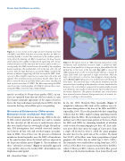
Figure 3. A cross section of the organ of Corti showing inner hair cells (IHCs) and OHCs with their stereociliary bundles. An OHC ste- reociliary bundle is circled. The central axis of the cochlear spiral is to the left of the drawing. An IHC is located over the bony (osseus) spiral lamina and is tightly enveloped with supporting cells. Sound- evoked vibrations will be reduced at this location relative to those oc- curring under the OHCs located nearer the middle of the compliant basilar membrane. The mottled dark circle in the hair cells is the cell nucleus. Note the large fluid spaces around the OHCs created by the absence of adjacent supporting cells that surround the IHCs. Audi- tory nerve fibers (eighth cranial nerve) contact hair cells at the end opposite their mechanosensory stereociliary bundle. More than 15 fibers contact the base of each IHC while a few course laterally and each contacts more than 15 OHCs. The fibers innervating the IHCs come from type 1 spiral ganglion cells (SGC1), whereas those inner- vating the OHCs come from type 2 SGCs (SGC2).
muscle) are within 10-30 nm of one another. OHCs, in con- trast, are separated from adjacent cells by as much as 1 μm. The elegant colonnade appearance of the organ of Corti is due to the large and unique spacing between OHCs, but the reason for the large extracellular spaces was puzzling.
Structural Evidence for Differences Between Inner and Outer Hair Cells
The invention of the electron microscope (EM) in the ear- ly 20th century provided a powerful new tool to examine the fine structure of cells because it could observe objects that were smaller than the wavelength of light. By midcen- tury, EM investigations had revealed structural similari- ties between all hair cells and revealed unique specializa- tions in OHCs. One of these was the presence of flattened membrane-bound organelles near the OHC cell membrane at the same location where the lateral wall was exposed to the large extracellular spaces (Figure 3). The membranes of these “subsurface cisternae” (Figure 4) invariably appeared crisp and flat, whereas the nearby OHC cell membrane was diaphanous and rippled.
22 | Acoustics Today | Spring 2017
Figure 4. The apical end of an OHC showing organization of the membranes and cytoskeletal structures. Left: a cylindrical OHC has been sliced parallel to its long axis along a plane defined by the dashed line in the insert at the top right. Top right: a view of the OHC looking down on the stereocilia bundle that has a typical W shape. Each small circle represents a single stereocilium, while the large circle represents a structure that disappears during develop- ment. Bottom right: high-power view of the lateral wall showing its three layers. The outermost layer is the plasma membrane and the innermost layer is made up of the subsurface cisterna. Sandwiched in between is the cortical lattice composed of circumferentially oriented actin filaments and axially oriented spectrin (spectrin filaments are the thinner filaments). The plasma membrane and cortical lattice form nanoscale motor elements that generate force at acoustic fre- quencies, resulting in electromotility.
In the late 1960s, Heinrich Hans Spoendlin (Figure 5) completed a laborious EM study of the auditory nerve fi- ber innervation pattern at the base of the IHCs and OHCs (Spoendlin, 1966). The results provided the first compelling evidence that the OHCs might be doing something entirely different than the IHCs. He meticulously reconstructed the auditory nerve fiber innervation pattern at the base of both the IHCs and OHCs by examining hundreds of ultrathin serial-section electromicrographs per hair cell. Each audi- tory nerve fiber that enters the organ of Corti arises from a single cell located in what is called the spiral ganglion (located closer to the spiral axis of the cochlea). He found that 90-95% of the fibers came from a population of spiral ganglion cells (SGCs) that he called type 1 SGCs (SGC1). The remainder were smaller fibers arising from type 2 SGCs (SGC2). SGC1 fibers were connected exclusively to the IHCs (about 15 fibers per IHC), whereas each SGC2 fiber passed radially near the basilar membrane, then turned basely and
