
Page 31 - WINTER2019
P. 31
thresholds that are six times smaller than those of nonmusi- cians (Micheyl et al., 2006).
Although different harmonics with the same f0 can have dif- ferent timbres, they nevertheless share the same sensation of pitch. In the most extreme case, a pitch can still be heard even when there is no energy at the f0 altogether as long as energy is present at multiple harmonics. This phenomenon, known as virtual pitch, has been both a challenge and a test case for neural models of pitch coding. Specifically, a neural mechanism that codes for pitch must yield an output similar to the f0 even when no energy is present at the f0 as long as the harmonics of the f0 are present.
Recording from the auditory cortex of marmosets (Callithrix jacchus), Bendor and Wang (2005) found that a small popu- lation of neurons in the anterolateral border of the primary auditory cortex responded selectively to both the f0 and har- monic complexes of overtones above the f0, thus providing a neural correlate of pitch constancy. Follow-up studies showed that this pitch constancy is likely accomplished using a com- bination of spectral and temporal cues. By building on these cues and then recombining pitches hierarchically to form melodies and harmonies, the neural coding of pitch provides the basis for the higher order coding of musical structure in the brain.
Amusia
One way to understand how pitch processing works in the brain is to look into individuals who have impairments in pitch-processing ability. Congenital amusia, also known as tone deafness, is a lifelong musical disorder that prevents individuals from developing skills in pitch perception and production despite no apparent deficits in speech, hearing, or cognitive ability (Ayotte et al., 2002). It is most commonly identified by using a neuropsychological test known as the Montreal Battery for Evaluation of Amusia (MBEA; Peretz et al., 2003). People with congenital amusia, who could not consciously report the directions of pitched intervals, could nevertheless produce (by singing) pairs of tones with above- chance accuracy in pitch interval direction (Loui et al., 2008). This dissociation between perception and production ability suggests that that there may be multiple paths toward audi- tory processing.
Studies from multiple neuroimaging methods have shown differences in auditory as well as auditory-motor brain pro- cesses that are linked to congenital amusia. Using structural
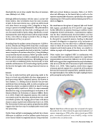
MRI and cortical thickness measures, Hyde et al. (2007) observed differences in the frontal lobe as well as in the superior temporal lobe of amusics, specifically in the superior temporal gyrus (STG) and the inferior frontal gyrus (IFG) as shown in Figure 2.
The simultaneous disruption of temporal lobe and frontal lobe regions, specifically the STG and IFG, may suggest further difficulties in memory, learning, or auditory-motor integration of pitch information. A parsimonious explana- tion for these simultaneously observed deficits was that white matter connectivity between the STG and IFG could be disrupted in congenital amusics, leading to abnormal neuronal development or migration at the end points of this connection. This was observed in a diffusion-imaging study in which the arcuate fasciculus, which connects the temporal and frontal regions of the brain, was smaller in volume in people with congenital amusia (Loui et al., 2009).
Other studies have employed MEG in combination with structural MRI to provide further support for a right fronto- temporal deficit in connectivity (Albouy et al., 2013), whereas ERP work suggests that congenital amusia may be fundamen- tally an issue with the lack of awareness of pitch information (Peretz et al., 2009). Taken together, results from congeni- tal amusia provide support for a crucial role of the pathway between the frontal and temporal lobes, probably mostly in the right hemisphere, that enables the tight coupling between pitch perception and production.
Figure 2. Model brain with brain regions that correspond to the regions discussed in the text. Colors indicate the approximate landmarks: red, superior temporal gyrus (STG); yellow, inferior frontal gyrus (IFG); green, ventromedial prefrontal cortex (vmPFC); orange, nucleus accumbens; blue, arcuate fasciculus.
Winter 2019 | Acoustics Today | 31
