
Page 63 - Fall2020
P. 63
lessons that started before age seven, while “nonmusi- cians” cannot receive more than one year of formal music training anytime in their lives. A second type of study that researchers later adopted is called longitudinal stud- ies, in which researchers follow a group of people as they begin to engage in music training for a period of time and observe changes in their brain compared with a group of controls who do not engage in music learning.
Most studies in the literature belong to these two categories, and we now review them in detail. From cross-sectional and longitudinal studies, three areas of neural differences emerged between trained and nontrained individuals, namely, neural structure, neural function in processing music, and neural function beyond music processing.
Neural Structural Differences
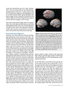
Researchers have found differences between musicians and nonmusicians in the structures of the brain. The differences have been observed in both the cortical gray matter and white matter characteristics. The gray matter of the cortex mainly consists of cell bodies. Specific brain functions, such as sound processing, are considered to be related to the gray matter characteristics in specific brain regions, such as the auditory cortex. One main charac- teristic of the gray matter is its volume or how much gray matter there is. Gaser and Schlaug (2003) examined the gray matter volume across all brain regions using MRI technology in three groups of participants: nonmusi- cians, amateur musicians, and professional musicians. Although both professional and amateur musicians sat- isfied the selection criteria for musicians, professional musicians further satisfied criteria that they were music performers or teachers who practiced for at least one hour a day while amateur musicians were people who played regularly but worked in fields outside of music. Researchers found gray matter volume differences across the three groups of participants in three cortical areas: auditory, motor, and frontal regions (Figure 1). Specifi- cally, professional musicians had the largest gray matter volume, whereas nonmusicians had the smallest volume in these regions. The differences observed support the idea that music learning involves strenuous motor prac- tice in addition to hearing and that this may result in bigger volumes in motor related regions. In fact, Bangert and Schlaug (2006) later found that professional musi- cians had increased gray matter volumes in the cortical regions specifically related to hand function. Moreover,
pianists showed a larger volume for the right hand while violinists showed a larger volume for the left hand, reflecting the differences in the hand dominance in play- ing these instruments.
On the other hand, cortical white matter generally con- sists of axons of neurons and its characteristics, such as the amount of white matter and the alignment of axons, generally reflect the “interconnection” or “communi- cation” across regions of the brain. For example, more white matter and better axon alignment support faster communication across brain regions. Bengtsson and colleagues (2005) compared a group of professional con- cert pianists with age-matched nonmusician controls in terms of their cortical white matter characteristics. The investigators found differences between the two groups in the axon alignment in the major pathways that con- nect the left and the right sides of brain, the frontal and the temporal regions of the brain, and the brain and the
Figure 1. Reconstructed cortical surface of a human brain from raw magnetic resonance imaging (MRI). Top: top of the brain with front of the head to the right. Bottom: right (R) and left (L) sides of the brain with the front of the head toward the center. The colored areas on the cortical surface indicate that there is a significant relationship between the gray matter volume of that region and an individual’s music training background. The brighter the color, the more robust the relationship. These regions are within the boundaries of the auditory, motor, and frontal cortices. Adapted from Gaser and Schlaug, 2003, with permission; copyright 2003 Society for Neuroscience.
Fall 2020 • Acoustics Today 63
