
Page 24 - Spring 2006
P. 24
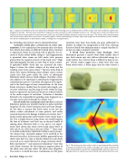
  Fig. 7. Sequence of simulations of the stress induced by a lithotripter shock wave (Fig. 2) in a cylindrical stone as shown in Fig. 6. The wave, modeled as a plane wave, propagates to the right. The linear elastic model shows compression as blue and tension as yellow and higher tension as red. The shock wave in water encircling the stone
37
reinforces the shear wave generated at the proximal corner of the stone and yields the highest maximum tensile stress in the stone.
well with the location where these stones fracture. The model, together with an understanding of cavitation, offers useful insight into the mechanisms of stone comminu- tion and has enabled progress in improving the complex, evolving process on fragmentation.
The location of the maximum agrees
 ber of findings of practical value to clinical lithotripsy.
Cavitation clouds play a critical role in stone com- minution: In vitro experiments (we mention only a few here) have shown that stones are difficult to break when cavitation is suppressed. Stones do not break well in glycerol, the vis- cosity of which softens bubble collapse,24 and fragmentation is prevented when stones are held under static pressure
25
The force of the fluid or shock wave impact following cloud collapse creates surface cracks that then grow under the stress of subsequent lithotripter shock waves or cloud collapses. Therefore, cavita- tion appears to be important in initiating the fragmentation (comminution) process. Cavitation also appears to be impor- tant in the final stages of stone comminution. As the stone breaks into pieces smaller than the shock pulse length, con- structive interference and focusing of waves within the frag- ments is suppressed, and internal stresses are reduced relative to the surface impact of cavitation. Cavitation is therefore believed to be responsible for grinding stone fragments to a
27
research showed severe damage to kidneys at extreme rates. Until recently, physicians rarely treated at rates slower than 2 Hz. This is largely because using slower rate would lengthen the time of treatment, and there was no obvious benefit of slowing down. However, both in vitro and in vivo experi- ments have shown that shock waves break stones better at
29-31
wave and act as nuclei that lead to a large cavitation cloud. Both experiments13,33 and numerical simulations34 show that such clouds attenuate the low-frequency negative phase of the shock wave and may reflect the shock wave altogether. The result is to shield the stone. Calculations of 2-Hz pulse
High speed photography of stones in water (Fig. 5) shows a dense- ly populated bubble cloud that can surround the stone. Figure 6 shows the violent collapse of the cloud and the
26
greater than the negative pressure of the shock wave.
resulting crater in the stone surface.
size that can be passed through the urinary tract.
Rate of shock wave treatment can be too fast: In clinical lithotripsy, patients are typically treated at a pulse repetition frequency of 2 Hz. Faster treatment rates have been attempt- ed, but there is concern that shock waves at fast rate can induce cardiac arrhythmias in some patients, and earlier
28
These stud- ies include model stones implanted in kidneys and a prospec- tive clinical trial. Cavitation appears to be involved in the observed rate effect. At faster rates, bubbles generated by one shock wave have less time to dissolve before the next shock
32
slow rate (0.5 or 1 Hz) than at fast rate (2 Hz).
 repetition rates show that clouds can grow sufficiently in number to collapse less energetically as well. Thus, reducing the rate of shock wave administration is a simple step that cli- nicians can take to improve treatment.
A broad focus promotes stone breakage: Most
lithotripters generate a narrow focal width smaller than 8
mm. Such narrow spot sizes deliver substantial energy to a
small volume, but a narrow beam is difficult to keep on tar-
35
get. Recent studies suggest that a wider focal zone may
break stones better. A beam larger than the stone creates a
   22 Acoustics Today, April 2006
Fig. 8. The fracture grows from surface cracks as may be generated by cavitation to the point of maximum stress. Cracks in this figure were etched 2 mm from the distal end and yielded a conical break. Collapsing bubble clouds have been seen in high-speed movies to ring the stone near the distal end where fracture first occurs.
