
Page 23 - Spring 2006
P. 23
but the negative pressure (rarefaction) phase preceded the positive phase of
15
16
measurable lesion. Delivery of stag-
gered dual pulses can be used to manipulate cavitation, and when the trailing pulse is timed to interrupt the cavitation cycle initiated by the first
17,18
Shear has the potential to damage cells: In vitro studies have shown that in the absence of cavitation, shock waves still generate differential forces (shear) and accelerations capable of
19
tearing cell membranes. Shear is
amplified by narrowing the beam or decreasing the shock rise time by increasing the shock strength. Shear damage to tissue by lithotripter shock waves has not been demonstrated in vivo, but it has been hypothesized that the structural inhomogeneities of organized tissues should act as foci to disrupt shock wave propagation, creat- ing local stress gradients sufficient to
20
cause mechanical failure. It may be
that shear stress tears vessels, causing blood to pool, and then cavitation takes hold, causing further damage.
The kidney vasculature shows a vasoconstrictive response to shock wave treatment: One of the most fas- cinating observations to come from studies of SWL trauma is the finding that shock waves stimulate blood ves-
21
sels in the kidney to constrict. The
induction of vasoconstriction proves to
be a physiologic response of consider-
able importance. Researchers have
shown that treatment of kidneys with a
minimal dose (~100 pulses) of low
energy shock waves acts to protect the
kidney from injury when a complete
dose of 2000 pulses is delivered at high
22
energy. That is, a priming dose of
shock waves capable of inducing vaso- constriction will protect the kidney from subsequent damage. In this way, strategically delivered shock waves can be used to minimize the adverse effects of a full clinical dose of pulses. As a first step, this suggests a protective strategy,
14
tiated by the negative phase was sup-
the wave.
Thus, bubble expansion ini-
Kidneys treated with conventional waveforms suffered a substantial lesion, while kid- neys treated with pulses produced by the pressure-release reflector (pulses that suppressed cavitation) showed no
pressed by the positive spike.
pulse, renal injury is reduced.
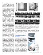
Fig. 5. Cavitation bubble clouds contribute to stone breakage in SWL. Top plate shows a cluster of bubbles that have formed at the leading edge of a gypsum model stone 100 μs after the passage of the shock wave. Middle plate illustrates the bubble cloud cycle caught in 100 μs steps, as shown using backlighting (top row) and incident light- ing (bottom row). The bubble cloud grows to fully engulf the end of the stone then collapses to a narrow spot. Cluster collapse can generate secondary shock waves (not shown) equal in amplitude to that of the incident pulse. Bottom plate shows several steps of the cloud cycle in which bubble activity appears to have forced open a crack
26 in the stone. (Reprinted with permission from Journal of Endourology. )
research to determine how shock waves break stones has yielded a num-
a recommendation that urologists start at a low power setting before increasing the power in order to give the kidney a chance to develop a self-protective response.
Progress in understanding stone comminution
Stone-free rates (a clinical metric
for relative success of treatment) for
current lithotripters are as much as 2-
3 times lower than values reported
with the first lithotripter (Dornier
HM3) used widely in clinical prac-
9,10
There has been considerable effort, therefore, to understand how stones break in order to improve these success levels, and
Roughly half of current treat- ments do not leave the patient stone- free, and as reported above no lithotripsy is without risk of side- effects. The original lithotripter is still considered the “gold standard” because of its low re-treatment rates, high stone-free rates, and low occur-
23
tice.
rence of side effects.
Fig. 6. High speed images of the cavi- tation cloud col- lapse and damage induced by SWL on the proximal face of a model stone. (Reprinted with permission from Journal of
26 Endourology. )
Progress in Lithotripsy Research 21
