
Page 21 - Spring 2006
P. 21
Progress in research on tissue injury in SWL: Making good use of acoustic principles
Most anyone with a background in acoustics would not be surprised to learn that lithotripter shock waves can cause tissue injury. That is, it seems reasonable to expect that a shock front moving through the urine space and the sur- rounding renal tissue and blood vessels could generate sig- nificant acoustic cavitation and focal shear stress, and that shock pulses capable of shattering stones might also have the potential to damage living cells. However, at the time that shock wave lithotripsy was introduced and, indeed, throughout much of the history of clinical lithotripsy, the medical community has been somewhat reluctant to accept that such adverse effects occur. In recent years, however, researchers have made considerable progress in characteriz- ing shock wave injury, including the mechanisms of shock wave action involved. Thus there has been a change in awareness of the potential for shock waves to cause trauma, and a new appreciation for the role that acoustics plays in understanding how shock waves break stones, the origins of tissue trauma, and what needs to be done to make lithotrip- sy safer and more effective. This work has generated practi- cal recommendations, specific steps aimed at improving clinical outcomes.
Tissue is responsive to shock waves, and shock wave
dose can be excessive: Shock waves may be intended to
break stones but, unfortunately, can cause collateral tissue
damage as well. Indeed, all SWL patients suffer some level of
tissue injury, and some patient groups, such as children and
the elderly, are at greater risk that this damage can be signif-
6
icant. Studies have shown that the lesion produced in
lithotripsy is acute vascular trauma in which the hemor-
7
rhage can be mild to severe. The lesion volume is dose-
dependent: more shock waves or higher amplitude pulses
cause greater injury. Also, hemorrhage leads to scarring that
8
in turn can lead to a permanent loss of functional tissue. In
addition, the long-term effects of SWL trauma can include hypertension, diabetes, and with multiple treatment ses- sions, a progression in stone disease to a type (brushite dis-
9
ease) that is significantly less responsive to SWL. This is
compelling evidence to minimize shock wave exposure, and to find treatment strategies that improve the efficiency of stone comminution.
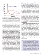
Fig. 2. Lithotripter waveforms generated by electrohydraulic and electromagnetic shock sources. Pressure waveforms measured in water were generated by the origi- nal electrohydraulic clinical lithotripter (red: Dornier HM3) and by a recent popu- lar electromagnetic lithotripter (blue: Storz SLX). All lithotripters produce a shock pulse consisting of a positive spike followed by a negative trough. The pulse of the electromagnetic lithotripters (EML) includes a trailing positive pressure oscillation.
source has been enclosed within a water-filled cushion, and the enclosing, acoustically-matched membrane (latex or silastic rubber) is coupled to the patient’s skin with gel or oil. Fluoroscopy remains the standard for targeting, especially in the United States, although many lithotripters have B-mode ultrasound as well. Shock wave sources have been engineered from three technologies (Fig. 1), which is evidence in itself that there is no consensus on correlation between source technology and performance. Electrohydraulic lithotripters (EHL) are the least complicated to manufacture, and use a spark source to generate the shock wave that is focused by an ellipsoidal reflector. Disadvantages are that shock strength tends to be variable, especially as the electrodes wear, and electrodes must be periodically replaced. In electromagnetic lithotripters (EML), a high current in a coil abruptly dis- places a plate to create an acoustic wave that is focused by the curvature of the plate, a lens, or a reflector. EML devices pro- duce stable and reproducible shock waves, and a well-built shock source has a lifetime of a million or more pulses. Currently, the three largest manufacturers of lithotripters provide only electromagnetic devices. In piezoelectric lithotripters (PEL), piezoceramic elements excited by a volt- age spike rapidly distend to produce an acoustic pulse that is focused by the curvature of the element or the scaffold sup- porting the elements. Like electromagnetic lithotripters, PELs rely on nonlinear acoustic propagation (see Anthony Atchley’s article in the first issue of Acoustics Today) to devel- op a shock wave, while the spark-generated pulse in EHL is shocked from inception. A potential advantage of PEL is the possibility of tailoring the pulse, altering the standard lithotripter waveform by changing the excitation of the ele- ments. Waveform shaping is just one of many areas where basic research has the potential to deliver significant improvements.
Ed.—The authors point out that a recent New York Times arti- cle (“Blasting of Kidney Stones Has Risks, Study Reports” by Lawrence K. Altman, 10 April 2006) cites a report* that, after nineteen years of follow-up study, shows that patients who received lithotripsy developed diabetes at almost four times the rate of those whose kidney treat- ment was other than lithotripsy. There was also a positive relationship between lithotripsy treatment and an increase in high blood pressure.
* A.E.Krambeck, et al., “Diabetes Mellitus and Hypertension Associated with Shock Wave Lithotripsy of Renal and Proximal Ureteral Stones at Nineteen Years of Follow-up.” J. Urology 175, 1742–1747 (2006).
Progress in Lithotripsy Research 19
