
Page 42 - January 2006
P. 42
Biomedical Ultrasound
Continued from page 38
imaging of deep tissues. Today, modern ultrasound scan- ners use the linear superposition of spherical or cylindrical wavefronts of arrays of tiny piezoelectric elements to pro- duce waveforms that can be steered or focused based upon the timing of element excitations. Beam forming can occur in both the emitted and received waveforms, and there is a great deal of engineering that goes into obtaining the images of, for instance, the fetus that may be the par-
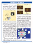
Fig. 3. If A-mode amplitudes are represented by the brightness of dots on a phos- phor screen and the transducer is scanned across the tissue, a B-mode (bright- ness) image results. Assuming a constant speed of sound allows quantitative imaging of deep tissues.
ents’ first glimpse of a new child. Nevertheless, the basic image is formed via sonar techniques not too much differ- ent from those employed in the 1960s.
Of more recent interest are techniques that rely upon the essential nonlinearity of human tissue as a propagation medium (see Atchley’s Nonlinear Acoustics Primer, Acoustics Today, pp. 19-24, October 2005). Figure 4 shows a typical imaging pulse from one element of an array trans- ducer. If an array element is excited at the same time with the inverse of this signal, the two pulses will mostly cancel each other’s fundamental frequency in the focal region. Tissue nonlinearities that give rise to waveform distortion, however, will result in the moving of energy from the fun- damental frequency to the second harmonic (twice the fundamental frequency). The second harmonic compo- nents will not cancel, but rather add together, producing a pulse with spatial resolution twice that of its B-mode equivalent and with improved signal-to-noise. This tech- nique is called tissue harmonic imaging, and most ultra- sound scanners in hospitals today transmit signals at one frequency but “listen” to the returning echoes at twice that frequency.
The ability of the essential nonlinearity of tissues to produce waveform distortion gives rise to sum and differ- ence frequencies when two frequencies overlap in space and time. Since the nonlinearity parameter known as B/A
Fig. 4. Addition of pulses with their inverse would cancel entirely if it were not for nonlinear addition of second-harmonic (and other) components.
varies with greater dynamic range in soft tissues than do acoustic impedance mismatches, a method of imaging this nonlinearity may provide important additional clinical information. To accomplish nonlinear imaging, one can measure the strength of the sum frequency or difference frequency by scanning a point of intersection of two dif- ferent-frequency pulses around the tissue volume.
A main limitation is the requirement to have large- amplitude, highly localized pulses intersecting within the tissue. Recently, a technique known as Time Reversal Acoustics (TRA) has provided a new tool to accomplish this goal. In TRA, an acoustic source located within the tissue produces a pulse that propagates along many different rays to a receiver, producing a complicated impulse response at the receiver. If the pattern of pulse arrivals at the receiver is then time-reversed, and the receiver now acts as a transmit- ter, the pulses follow the same rays in reverse, coinciding at the original source location as a highly spatially and tem-
Fig. 5. Time-reversed signals sent from each transducer coincide at a common location in space and time, allowing generation of sum and difference frequen- cies that can be used to image tissue nonlinearities.
40 Acoustics Today, January 2006
