
Page 43 - January 2006
P. 43
Biomedical Ultrasound
porarily focused pulse. In effect, the time-reversed signal at the receiver acts as an optimum filter to deliver sub-wave- length focusing at the (original) source location.
In practice, TRA requires that a source be placed in the body, although this source could be the tip of a biop- sy needle used to reflect incident ultrasound. If the receiver is not one, but many (reciprocal) transducers mounted on a surface, a highly focused spot can be gen- erated at the site of the original source. For imaging the nonlinear parameter of tissues, two resonators may be used, each holding reciprocal transducers of either fre- quency f1 or f2. If Time-Reversal Acoustics techniques are used to place the focal spot of both resonators at the same spatial point, the sum or difference frequency due to the nonlinear interaction of these two waves can be meas- ured as seen in Fig. 5.
Due to the TRA focusing of both frequency signals, the resulting large amplitudes allow “synchronized stir- ring” of f1 and f2. The inherent nonlinearity of tissue pro- duces sum and difference frequencies. The sum or differ- ence signal can be filtered and detected by a probe, and the
location of the coincident spot moved to a new location to image the rest of the tissue volume. The amplitude of the signal is proportional to the tissue nonlinearity parameter at each point.
In conclusion, modern ultrasound images rely upon tissue nonlinearities to produce clearer pictures of tissues. New techniques are emerging that make even greater use of nonlinear acoustics for imaging and therapy.
Acknowledgments: The author would like to thank Pierre D. Mourad and Shahram Vaezey (University of Washington, Seattle, WA), Armen Sarvazyan (Artann Laboratories, NJ), Christy Holland (Univ. of Cincinnati, OH), and Mickael Tanter (Laboratoire Ondes et Acoustique, ESPCI, France) for use of their figures.
E. Carr Everbach, Associate Professor of Engineering at Swarthmore College, is the former chair of the ASA Biomedical Ultrasound/Bioresponse to Vibration technical committee. This article is based on his tutorial lecture present- ed at the 150th ASA meeting in Minneapolis.
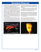
Fig. 6. Doppler scan of an artery showing blood flow.
Fig. 7. Ultrasound reconstructed image of a baby’s face in the womb
Echoes 41
