
Page 14 - Summer 2008
P. 14
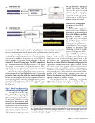
 Fig. 7. Plane wave impinging on a 2-element hydrophone array: Frequency and bearing estimation problem: (a) classical spec-
tral (temporal and spatial) estimation approach; (b) Model-based approach using parametrically adaptive (nonlinear)
18,19
processor to estimate bearing angle, temporal frequency and the corresponding residual or innovations sequence.
we have discussed or time/space varying (e.g., ocean) where we construct the processors using recursive-in-time or recursive- in-space techniques or both to capture the ever-changing medi- um or motion of the acoustic problem that must be solved.
Model-based tomographic imaging for biomedical imaging
Tomographic imaging is governed by partial differential propagation equations evolving from a full-field wave model to
20,21
It can be thought of as a methodology of solving an inverse problem. Consider the development of a tomographic image of breast tis- sue to determine if a cancerous tumor is or is not present. This technology is based on Fourier imaging techniques and diffrac- tion tomographic reconstruc- tion of the tissue using multiple scans through the breast (object). Fourier imaging essentially obtains projections in object space using Fourier transforms (1 dimensional) to “fill in” regions in the 2 dimensional (2D) Fourier space and is based on the Fourier diffraction theorem equating a projection (plane wave propagation) in object space to a region (arc) in 2D-Fourier space. Once the space is filled, a 2D inversion is performed using the model to perform a backpropagation similar to a numerical time-reversal to reconstruct the object and create the image. This is similar to computer-aided tomo- graphic (CAT) reconstruction employing x-rays using the Fourier slice theorem along a line, but here the acoustic prop-
generate its solutions.
20,21
The application of this model-based (distributed)
agation is modeled by the wave equation.
22
observe the usual ultrasonic reflection image (not tomogra-
approach to the breast tissue is shown in Fig. 8.
Here we
 more computationally intensive than the spectral estimators used in the classical approach; however, the results are appealing as shown in Fig. 7b. We see the bearing angle and temporal fre- quency estimates as a function of time converging to the true values of 50 Hz and 45o bearing angle. The MBP also produces the “residual or innovations” sequence, (shown in the figure) that is used in determining its overall performance for valida- tion. In this case the sequence must be statistically zero-mean
7
and white (uncorrelated) for optimal performance.
Thus, the classical approach simply performs spectral esti- mation temporally and spatially (beamforming) to extract the parameters from noisy data, while the model-based approach embeds the unknown parameters into its propagation, measure- ment, and noise models enabling a solution to the joint estima- tion problem. The performance of the MBP is validated by ana- lyzing the statistics of its innovations sequence. This completes
the application.
Step 5: Model-based processing (distributed physical approach)
This step can be the most com- plex depending on the embedded model representation. Typically, the phenomenology is represented by a set of partial differential equations characterizing the propagation medi- um and a set of noisy measurement equations that still can be character- ized by a state-space representation in some form or another. Whether the application is based on time-invariant statistics like most of the applications
  Fig. 8. Ultrasonic diffraction tomography is a model-based method based on the wave equation propagation model and backpropagation techniques to construct a meaningful reconstruction. The model-based tomographic reconstruction shown on the far right image compares quite well to the usual ultrasonic scanner shown on the far left (reflection ultra- sound) and the x-ray CAT scan (middle).
 Signal Processing in Acoustics 13
