
Page 48 - Volume 12, Issue 2 - Spring 2012
P. 48
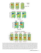
  Fig. 5. (Top row) Comparison of a pre-behavior, normalized quiescent spatio temporal STRF (left panel) and behavioral reference field (STRF) (middle panel). Color scale: increased (red) to suppressed (blue) firing about the (green) mean; red and blue are statistically significant deviations from the mean. Top row--black arrow: frequency of the target tones during the detection task. The difference between the normalized quiescent and detection STRF is shown in the right panel (STRFdiff). Asterisk marks the location of maximal change. Second and third rows--facilitative STRF plasticity in AI. STRFs from two single-units in AI illustrate typical changes observed during per- formance of the tone-detection task. Second row—pre-behavior STRF (left panel). Localized enhancement of an excitatory region in the STRF at the target frequency dur- ing behavior (middle panel). The post-behavior quiescent STRF (right panel) reverted immediately to a RF very close to its original shape. Pre-behavior STRF (left panel). Local decrease (near elimination) of lower inhibitory sideband at the target frequency in the detection STRF (coincident maxima at target frequency). Fourth Row--three quiescent STRFs interleaved with two sequential two-tone discrimination tasks, all measured during recordings from the same neuron. The times at which STRFs were meas- ured relative to the beginning of recording are shown on top of each panel. The arrows mark the reference (green) and target (red) frequencies used. Note the disappearance of the excitatory area in the near 250-Hz in the STRFs measured during the discrimination tasks. Reprinted with permission from Fritz et al. (2005b).
Auditory Cortical Function 47
