
Page 31 - Volume 8, Issue 4 - Winter 2012
P. 31
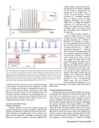
  Fig. 4. (Top) Representative focal pressure waveform used for histotripsy. The pulse is initially a sinusoidal tone burst, but at the focus it is distorted by the combined nonlinear propagation and diffraction effects to produce the asymmetric waveform with higher peak positive pressure (p+), lower-amplitude peak negative pressure (p-), and high-amplitude shocks formed between negative and following positive phase as shown in the inset frame. (Bottom) Pulse-periodic timing schemes for two forms of histotripsy. The blue sequence shows the cavitation- cloud histotripsy scheme with microsecond-long pulses applied at 100-1000 Hz. The red sequence shows the boil- ing histotripsy scheme, employing millisecond-long pulses at a rate of 0.5 - 1 Hz.
 of shocks with this vapor cavity causes mechanical damage of tissue before thermal effects become significant. Usually, 10 to 50 pulses are sufficient to fractionate the focal vol- ume.15 In both pulsing schemes, ultrasound is only being delivered to the focus about 1% of the treatment time. The periods in which the transducer is not emitting give the tis- sue time to cool, thus preventing accumulation of heat and thermal damage within the tissue volume. In this way, a completely mechanical effect is preserved.
Cavitation cloud histotripsy
Acoustic cavitation
Acoustic cavitation occurs when bubbles expand and contract in response to a pressure waveform. At low pressure amplitude, this behavior is relatively mild and results in frac- tional oscillation in the bubble radius versus time. However, at higher pressure amplitudes, the bubble can undergo a huge expansion as tension is applied to the liquid, followed by a
which house tiny gas pockets.
Inertial cavitation thresholds in vivo can largely vary depending on the existence and distribution of
these nuclei.
Cloud cavitation in histotripsy
During histotripsy, many cavitation bubbles are expand- ed forming a dense cloud over the focal volume (Fig. 5). The cloud does not continuously increase in density as the acoustic pressure increases, but forms suddenly at a distinct pressure threshold. The value of this threshold, however, is about an order of magnitude larger than reported thresholds for inertial cavitation from single bubbles at 1 MHz. Interestingly, the formation of clouds is also probabilistic— that is, the cloud will not necessarily form on the first pulse applied above the threshold, but rather can take hundreds or thousands of prior pulses before a cloud will suddenly form, despite each pressure pulse being essentially identical. In this sense, cloud formation is “all-or-nothing.”12 When the pres- sure amplitude is sufficiently high (p- > 20MPa), the bubble
violent collapse as the mass of the mov- ing liquid forces the bubble to implode. As this motion is dominated by the momentum of the bubble wall, it is termed “inertial cavitation.” Inertial cavitation is a threshold phenome- non—it requires certain pressure amplitude, dependent on the initial bubble size, to undergo the explosive expansion. In cavitation-cloud his- totripsy, this large growth and collapse of bubbles creates a transient strain on the surrounding tissue, resulting in its fractionation.
The tensile pressure required to create the bubble nucleus for inertial cavitation in water is enormous—theo-
20,21
However, the threshold to expand a
pre-existing bubble in water or tissues is
lower; inertial cavitation can occur even
at modest pressures of p ~ 1 MPa at -
22-24
retically estimated at ~50 – 140 MPa.
Similarly, divers encounter decompression sick- ness through outgassing of bubbles in the blood after exposure to relatively small overpressures. These observa- tions suggest the body harbors small pre-existing gas nuclei which can be driven to undergo explosive growth. However, free bubbles are generally not stable by themselves—they tend to dis- solve over time due to the Laplace pres- sure created by surface tension of the liquid on the bubble. Instead, they are thought to be stabilized by different mechanisms such as crevices in solid particles or even macromolecules
25,41
ultrasound frequencies.
Disintegration of Tissue Using HIFU 27
