
Page 36 - Volume 8, Issue 4 - Winter 2012
P. 36
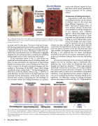
 Fig. 11. Boiling histotripsy lesions lesions produced in excised bovine liver by four independent exposures using a 2 MHz HIFU transducer of 4.4 cm diameter and focal length. Shock amplitude at the focus was about 70 MPa and time-to-boil was 4 – 5 ms.
 lesions with different degrees of ther- mal effects which can be controlled by varying parameters of the pulsing scheme.
Mechanisms of boiling histotripsy
Experimental studies have shown
that boiling histotripsy can be induced
in different types of soft tissues and
using different frequencies of 1 – 3
15
MHz. Mechanical fragmentation of
tissue without thermal denaturation was realized if several key components of the exposure were combined together: shock fronts higher than 40 MPa were present at the focus, shock amplitude was high enough to initiate boiling in several milliseconds, pulses were a little longer than time-to-boil, and the repetition rate of the pulsing
scheme was slow enough so that thermal effects did not
accumulate. No histotripsy occurred if any of these compo-
nents was absent. However, it was not clear how mm-sized
vapor bubbles can fragment tissue into sub-micron pieces.
Recently, it was proposed that the major mechanisms
involved in this process are acoustic atomization and for-
mation of a miniature fountain from tissue into the boiling
36,37
droplets into the air creating a fog is a well-known phe- nomenon that occurs when an ultrasound wave passes
38,39
hypothesis, the following scenario of boiling histotripsy
40
bubble.
Ultrasound atomization or the emission of small liquid
When high intensity ultrasound is focused at the interface, the acoustic radiation force pushes the liquid and creates a fountain. These effects have been used as a basis of air humidifiers and medical nebulizers. It was hypothesized that similar to liquids, tis- sues can be atomized and produce fountains. Based on this
through the liquid-air interface.
process was proposed and tested (Fig. 12).
High amplitude
  Fig. 12. Illustration of the proposed boiling histotripsy mechanism. (Left) Shock waves are superfocused and rapidly heat tissue to a boiling temperature in a small vol- ume at the focus. The boiling bubble is initiated in milliseconds and grows to a millimeter size. Shock waves interact with the vapor cavity generating atomization and an acoustic fountain from the tissue interface into the cavity. (Right) Experiment mimicking histotripsy processes in the vapor cavity. A frame of high speed photogra- phy showing atomization and a fountain at the free tissue/air interface generated by focusing the ultrasound beam at the surface of bovine liver. (Adapted from part of a figure in reference 40)
duration, and 0.01 duty factor. The time-to-boil was first pre- dicted using weak shock theory and then used to define pulse lengths and duty factors. The sequence of 50 pulses produced a liquefied cavity in the tissue. Histological studies also con- firmed that tissue thermal injury in such lesions is negligible compared to the mechanical injury caused by the exploding
34
No thermal damage was observed in the lesion content shown in Fig. 11. However, if pulse duration, the duty factor, or the focal pressures were increased, thermal denaturation of the lesion content was
34 detected both visually as blanching and with histology. It
was therefore shown that boiling histotripsy can produce
boiling bubble and its further interaction with shocks.
In the experiment shown in Fig. 11, time-to-boil was predicted theoretically and occurrence of boiling within each pulse was also controlled in the experiment. As large boiling bubbles are strong scatters, onset of boiling at the focus pro- vided a contrast for B-mode ultrasound imaging and also resulted in fluctuations of the HIFU source drive voltage
35,14
recorded by a high-voltage probe.
32 Acoustics Today, October 2012
