
Page 27 - 2017Spring
P. 27
What Powers Outer Hair Cell Electromotility? - The Silent Current
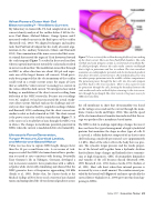
My laboratory in Gainesville, FL, had completed an in vivo current-density analysis of the cochlea before I left for Ge- neva. Paul Manis, Michael Zidanic, George Spirou, and I measured evoked currents in the fluid spaces of the cochlea (Brownell et al., 1983). We adapted techniques and analytic tools that Paul had developed for his study of neural orga- nization in the auditory brainstem (Manis and Brownell, 1983). Our examination of the inner ear revealed that acous- tic stimuli that moved the organ of Corti downward toward the scala tympani (Figure 7) resulted in decreased ion flow, whereas upward movements toward the scala media resulted in an increased ion flow. The modulation in ion flow through an OHC in either direction was about 500 pA and repre- sents one of the largest known cell currents. It had previ- ously been proposed that the electroanatomy of the cochlea could result in a steady current across the organ of Corti that we called the “silent current,” in analogy to a current in the retina called the dark current. We interpreted our initial findings as modulation of the silent current resulting from deflection of the OHC stereocilia. Because our recordings were AC coupled, we had not measured the actual steady- state silent current. Michael took on the challenge and sev- eral years later reported his DC-coupled recordings (Zidanic and Brownell, 1990) confirming that the silent current was similar to what we had estimated in 1982. The silent current is the power source for cochlear transduction (Figure 7). It is the source of a steady flow of ions through the OHC even in silence. The changes in membrane potential generated in OHCs when the current is modulated drive electromotility.
Ultrasonic Force Generation,
Turgor Pressure, Cortical Lattice,
and Membrane-Based Motor
Video was too slow to capture OHC length changes faster than the 30 per second frame rate. A succession of tech- niques revealed that OHC electromechanical force produc- tion occurred at rates consistent with hearing. Eventually, Tony Gummer’s lab in Tübingen, Germany, developed a way to measure isometric force production with a calibrat- ed probe while electrically stimulating and showed that the OHC generates force at frequencies approaching 100 kHz (Frank et al., 1999). Before that, Joe Santos-Sacchi at the Medical College of New Jersey used a variety of ion-channel toxins and manipulated the electrochemical gradients across
Figure 7. Cross section of the cochlea showing the flow of ions making up the silent current. There are three fluid-filled chambers. The scala vestibuli and scala tympani contain a conventional extracellular flu- id called perilymph, whereas the scala media contains endolymph that is high in potassium and low in sodium. The stria vascularis maintains an endolymphatic potential and electrochemical gradient that drives the silent current (arrows). The metabolically active stria vascularis pumps potassium into the middle cochlear compartment. The potassium passes through the hair cells into the scala tympani and then back to the stria vascularis. There is also a passive route for potassium through the cells, forming the boundary between the scala media and scala vestibuli before returning to the stria vascu- laris through the perilymph. The silent current is the power source for OHC electromotility.
the cell membrane to show that electromotility was based on the voltage across and not the current through the mem- brane (Santos-Sacchi and Dilger, 1988). This and the speed of the electromechanical transduction indicated that the en- ergy was produced by a membrane-based motor.
The OHC is able to undergo rapid shape changes because it does not have the typical arrangement of rigid cytoskeletal proteins that maintains the shape in other types of cells. It is, instead, a cellular hydrostat composed of an elastic outer shell enclosing a modestly pressurized core (approximately 1-2 kPa). Most of the other cells in the body have no pres- sure. The cytosolic turgor pressure and the tensile proper- ties of the lateral wall together form a hydraulic skeleton that facilitates shape changes and hydraulic force transmis- sion at high frequencies. OHC electromotility diminishes and vanishes if the cell becomes flaccid (Brownell 1983, 1990; Brownell et al., 1985; Santos-Sacchi, 1991; Shehata et al., 1991). Most cells burst when their internal pressure is increased by even a small amount. The reinforcement pro- vided by the lateral wall (Figure 4) and more specifically the cortical lattice (Oghalai et al., 1998) prevents this from hap- pening in OHCs.
Spring 2017 | Acoustics Today | 25
