
Page 41 - Fall2020
P. 41
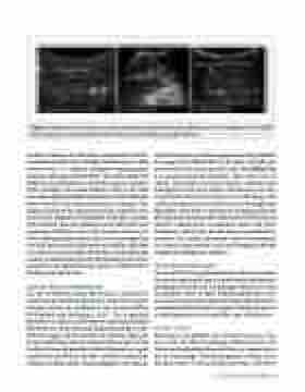
  Figure 5. Abdominal ultrasound. A: normal bowel. B: abnormal bowel with fluid-filled intestines and echogenicity of free fluid. C: abnormal bowel showing thickened bowl wall and edema (fluid) of the affected bowel.
to adults, neonates can suffer from complications of intesti- nal malrotation (abnormal twisting of the intestines), which, in some cases, is a surgical emergency requiring quick diagnosis and surgical intervention. The gold standard for diagnosis of malrotation is a study that requires transport to the radiology suite, liquid contrast placed in the stom- ach, and multiple plain film radiographies, all of which have inherent risks in the small and premature neonates. The pediatric literature has demonstrated the utility for ultra- sound in the diagnosis of malrotation with high sensitivity and specificity through evaluation of the blood flow and orientation of blood vessels to the intestines (Garcia et al., 2019). Abdominal ultrasound is also used to interrogate for free fluid and provide information on whether this fluid is anechoic (e.g., clear fluid), hypoechoic and nonhomoge- neous (e.g., blood or presence of stool indicating a tear in the intestine), or loculated fluid (e.g., abscess or blood within the lining around the liver).
Cardiac (Echocardiography)
The use of bedside cardiac ultrasound to characterize cardiovascular health and guide care of the sick newborn has now become the standard of care in many NICUs (El-Khuffash and McNamara, 2011). The recognized limitations of clinical and laboratory measures of normal blood flow out of the heart and adequate blood flow to the body has supported the need for an extensive approach to the monitoring of the neonatal cardiovascular system. Cardiac ultrasound provides detailed information regard- ing the function of the heart that cannot be obtained by clinical assessment alone. Echocardiography must include
an initial evaluation confirming normal anatomy, followed by an appraisal of blood flow to the lungs and body and assessment of the cardiac muscle health (see Multimedia 2 at acousticstoday.org/ruossmm). Three main uses of cardiac ultrasound in neonates include evaluation and management of neonatal cardiac transition from in utero to postbirth, elevated blood pressure in the lungs, and cardiac dysfunction due to infection or poor oxygen and blood flow to the heart. Utilization of emerging and serial physiological assessment by cardiac ultrasound may help identify cardiovascular compromise earlier and guide therapeutic intervention, thereby improving outcomes in neonates. The cardiac ultrasound serves to complement the clinical examination, with serial imaging offering insights into therapeutic response.
Procedural Ultrasound
The use of POCUS to guide invasive procedure in neonates has grown significantly over the past decade, and includes obtaining vascular access and drainage of fluid or air from the abdomen, heart, or chest. Additional procedure, such as monitoring of endotracheal (breathing) tube placement and removal of fluid from the spinal canal (also known as a lumbar puncture) are beyond the scope of this review.
Vascular Access
Placement of peripheral and central access into arter- ies or veins for close monitoring of blood pressure, lab draws, and medication administration is a common prac- tice in neonatology. Ultrasound-guided vascular access has been shown to decrease placement time, with fewer
 Fall 2020 • Acoustics Today 41
