
Page 40 - Fall2020
P. 40
POINT-OF-CARE ULTRASONOGRAPHY IN NEONATES
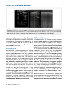
Figure 4. Lung ultrasound. A: normal lung in brightness mode (B-mode), showing acoustic shadowing from the ribs and a reverberation artifact from the pleural line. B: severe lung disease of prematurity, showing a rough appearing pleural line and coalesced comet tail artifacts. (B-mode). C: normal lung movement with breathing in motion mode (M-mode).
Lung ultrasound in neonates with delayed transition after birth consist of a normal pleural line, reverberation artifacts from the pleural line, few thin comet tails in the superior lung field (front of the chest), and increased com- pacted comet tails in the posterior lung field (back of the chest) (Raimondi et al., 2019).
Head Ultrasound
Head ultrasound is utilized for a noninvasive interroga- tion of structures within the skull through open fontanels (soft spot or spaces between areas of the skull) in newborn infants. Evaluation of the brain requires higher frequency transducers and the use of a far-field, wide-view sector transducer (Konofagou 2017). Occasionally, near-field evaluation is required, and a linear-array probe can be uti- lized. Indications to perform head ultrasound in a neonate include serial evaluation for bleeds around and within the brain and fluid collection within the brain. Intraventricular hemorrhage, which is bleeding within the ventricles or cen- tral areas of the brain that contain spinal fluid, is a cause of significant morbidity in neonates that are born early. As a greater proportion of extremely preterm infants are surviving in the NICU, there is an increasing need for tech- nologies to evaluate and monitor the evolution of these brain bleeds. The prompt identification of severe brain bleeding in the setting of acute clinical change can help guide management and modify decision making.
Abdominal Ultrasound
The rapid assessment for abdominal pathology with ultra- sound is recognized as a critical tool in adult and pediatric trauma patients and has recently become more widely uti- lized in neonates (Lynch et al., 2018). High-frequency linear or low-frequency curvilinear probes are utilized for abdomi- nal ultrasound, with the choice being based on the depth of structure and resolution needed. Plain film abdominal radiography (X-ray) is still the initial tool employed for evaluation of intestinal pathology and bowel health in neo- nates (van Druten et al., 2019), but it is limited by its inability to assess for malrotation (twisting of the bowel), bowel health, and abnormal fluid collections. Abdominal ultra- sound can interrogate for normal bowel health (Figure 5A) by evaluating for absence of intestinal movement or good blood flow, bowel wall thinning and thickening, fluid filled intestines, and echogenicity of free fluid (Figure 5B; Cuna et al., 2018). Recent experts and international consensus task forces have recommended abdominal ultrasound as a diagnostic modality for several abdominal emergencies in neonates and POCUS-guided paracentesis (bedside needle drainage of free fluid in the abdomen; Singh et al., 2020).
Abdominal ultrasound has been shown to be more sensi- tive in the early detection of necrotizing enterocolitis (van Druten et al., 2019), a severe life-threatening complication of preterm birth that causes bowel death (Figure 5C). Similar
40 Acoustics Today • Fall 2020
