
Page 38 - Fall2020
P. 38
POINT-OF-CARE ULTRASONOGRAPHY IN NEONATES
et al., 2011). Spatial resolution is divided into axial, parallel to the beam; lateral, perpendicular to the beam; and eleva- tion, reference to thickness of the slice of the images and similar to lateral resolution but in the orthogonal plane. Temporalresolutionistheabilitytodetectamovingobject and is mainly determined by the frame rate.
Ultrasound Artifacts
Ultrasound machines make assumptions in forming an image that does not always accurately show the structures or flow patterns being interrogated (Feldman et al., 2009). The more common artifacts in POCUS can be divided into beam artifacts, multiple echoes artifacts, velocity errors, and attenuation errors.
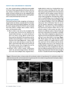
(1) Beam artifacts occur due to the assumption that the sound waves received by the transducer are located within the beam. As a result, side-lobe arti- facts occur when a strong reflector outside of the beam sends an echo back to the transducer and the machine inaccurately places a focus in the wrong location on the image (Figure 3A). A beam-width artifact occurs when the beam is wider than the object, echoes from other objects are returned, and the machine assumes these echogenicities are the same, altering the brightness of the image.
(2) Multiple echoes artifacts include reverberation, comet tail, and mirror image artifacts. Reverbera- tion occurs when multiple echoes originate from a
highly reflective surface (e.g., the pleural line-tissue connection between the lungs and the chest wall). The echoes go back and forth between the transducer
and reflector, and the machine assumes that these echoes are accurate and displays them as hyper- echoic (white) parallel lines. Reverberation artifacts can be a normal finding as seen in lung ultrasound with reverberation from the pleural line (Figure 3B) or an abnormal finding as seen in the abdominal ultrasound, with reverberation from free air out- side the intestines indicating a surgical emergency (Figure 3C). Comet tail is a type of reverberation, but instead of one reflective surface, there are two reflective surfaces. In this scenario, one echo, stuck between two reflective surfaces, bounces back and forth and is detected by the transducer as multiple echoes. This artifact is displayed as a hyperechoic tri- angle due to decreased attenuation of the later echoes and can be seen when fluid is in the lungs (Figure 3D). Mirror image occurs when the object being evaluated is adjacent to a highly reflective surface.
The sound waves hit the reflector and then hit the nearby medium and return to the transducer. The machine displays a duplicated image on both sides of the reflector due to the assumption that echoes are from two different mediums and can be seen when doing a lung ultrasound where the mirror image of the liver is visualized instead of the lung (Figure 3E).
Figure 3. Ultrasound artifacts. A: beam artifacts. B: reverberation artifacts in lung ultrasound. C: reverberation artifacts in abdominal ultrasound. D: comet tail artifacts from lung ultrasound. E: mirror artifacts in lung ultrasound. F: attenuation artifacts with acoustic shadowing with normal lung ultrasound.
38 Acoustics Today • Fall 2020
