
Page 36 - Fall2020
P. 36
FEATURED ARTICLE
Emerging Clinical Applications of Point-of-Care Ultrasonography in Newborn Infants
J. Lauren Ruoss, Catalina Bazacliu, Daphna Yasova Barbeau, and Philip Levy
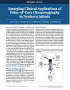
Point-of-care ultrasound (POCUS) is a bedside tech- nology with multiple emerging applications that can complement what is clinically suspected, thereby offering novel physiological insight into disease processes (Figure 1). POCUS has been used by nonradiologists for over 30 years in adult and pediatric care but has been slower to evolve in newborn medicine (Miller et al., 2019) even though critically ill neonates are some of the most vul- nerable patients that cannot be easily transported out of the neonatal intensive care unit (NICU). Thus, POCUS provides real-time diagnostic information at the crib side regarding clinical pathology and response to interven- tion, thereby enabling providers to more easily integrate the clinical examination findings with rapid and serial sonographic imaging (Conlon et al., 2019). POCUS is now being used by neonatologists for bedside assessment of lung pathology, heart function, intestinal health, brain bleeds, procedural guidance, and rapid identification for the etiology of acute clinical change. With a growing rec- ognition of the clinical utility of POCUS in the NICU (Miller et al., 2019), it is imperative to have a complete understanding of this technology. Accordingly, in this review, we discuss the physics of POCUS, the applications of POCUS in the critically ill neonate, and guidelines and limitations for wide use of this modality (Figure 1).
Physics of Ultrasonography
Understanding the basic physics principles of ultraso- nography as they relate to POCUS, including sound wave characteristics, ultrasound transducer mechanics, image optimization, and artifact recognition, is a prerequisite to enhancing image quality, improving diagnosis, and avoiding common errors. In this section, we review the common terminology and physics behind the guiding principles of ultrasonography.
Image Production
Medical ultrasound consists of using high-pitched sound (ultrasound) bouncing off tissues to generate images of inter- nal body structures. Ultrasound pulses are longitudinal waves commonly described by their frequency, wavelength, ampli- tude, and velocity. In medical imaging, there are two types of sound waves: (1) continuous, which is used in Doppler ultrasound and do not contribute to formation of an image, and (2) pulse, which generates images. Transducers equipped with polarized material inside the core, known as piezoelec- tric crystals, convert electrical current signals into mechanical vibrations through a tissue medium (such as tissue, fluid,
©2020 Acoustical Society of America. All rights reserved.
36 Acoustics Today • Fall 2020 | Volume 16, issue 3
Figure 1. Medical applications of point-of-care ultrasound in neonates.
https://doi.org/10.1121/AT.2020.16.3.36
