
Page 37 - Fall2020
P. 37
bone, or air) and compress when sound waves return from the tissue to the probe, changing the mechanical vibrations into an electrical signal to create an image (Groves et al., 2018).
There are three basic modes of electronic representation of pulse-wave sound that are generated with returning signals and displayed on a monitor.
(1)Amplitude mode (A-mode) provides informa- tion about the depth of the structures using the amplitude of the returning echo. A-mode is most commonly utilized with ophthalmology and was initially utilized with echocardiography.
(2) Motion mode (M-mode) is used to demonstrate motion across a single line of the ultrasound beam. It is commonly used to evaluate cardiac muscle movement to demonstrate strength of the muscle and effectiveness of the heart’s ability to pump blood to the body. M-mode is also used to evaluate for normal lung movement with breathing. Neo- nates that rapidly develop difficulties breathing may have a life-threatening accumulation of air in the chest that can be detected by the absence of normal lung movement on M-mode (Groves et al., 2018).
(3) Brightness mode (B-mode), the most common mode, produces a two-dimensional image where areas of different brightness (black, gray, and white) represent the sound waves (echoes) returning from the objects within the ultrasound beam. The signal returning from the body to the transducer is divided into pixels based on amplitude, ranging from black to white with ranges of gray in between (bright pixels represent high amplitude, such as bone; dark pixels represent low amplitude, such as blood).
Ultrasound Transducers
Curvilinear, linear (microlinear), and phased arrays are the most common types of transducers used in medical ultra- sound in neonates (Groves et al., 2018; Corsini et al., 2020). Linear and microlinear probes are high frequency, providing better resolution at superficial depths, and are commonly used in the evaluation of lung ultrasound and obtaining vas- cular access. Phased-array probes are high-frequency probes with a small footprint whose signals penetrate deeper into the body than the signal from linear probes but with pre- served resolution. They are utilized for head and cardiac ultrasound. Curvilinear probes are low-frequency probes with wide ultrasound beams that penetrate deep into the body and are utilized for abdominal ultrasound.
Ultrasound-Tissue Interactions
Ultrasound wave interaction with the mediums encoun- tered along its beam produces images (Gray et al., 2019). Sound energy is attenuated or lost because parts of it are absorbed, reflected, scattered, or refracted. In POCUS, the part of the scattering that does reach the transducer and generate images is called backscatter. The nature of the backscattered signal depends on the intrinsic properties of the tissue and it can be useful in evaluating a structure with different echogenicities (e.g., heart tissue). Echogenicity describes the ability of a surface to reflect an echo (sound wave) back to the transducer. In B-mode, echogenicity is represented on the image as anechoic (black), hypoechoic (gray), and hyperechoic (bright white).
Image Optimization
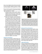
In POCUS, image optimization can be done in several ways including, for example, with the heart changing the output power (brightness), the receiver gain (brightness), the time gain compensation (offset attenuation with enhancement of image resolution), and the harmonics (improves the signal-to-noise ratio; Figure 2). These maneuvers assist in improving the spatial and temporal resolutions that are impacted by the ultrasound beam properties (Abu-Zidan
Figure 2. Image optimization. The gain buttons amplify the reflected signal in postprocessing. The time gain compensation controls the overall brightness of the image at different depths. Each slider controls gain selectively at a certain depth. A: sliders aligned. B: sliders adjusted with an increase in time gain compensation, resulting in a brighter image. RV, right ventricle; LV, left ventricle; RA, right atrium; LA, left atrium.
Fall 2020 • Acoustics Today 37
