
Page 28 - Spring 2006
P. 28
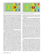
   Fig. 14. Predicted acoustic scatter of a lithotripter shock wave from an intact cylindrical stone (left) and a cylindrical stone fractured vertically through its center (right).
48
The differences can be detected by passive broadband remote hydrophones, and would give indication of whether the stone is fragmenting.
 scattering of the shock wave off the stone to detect a change in stone size. The same linear elastic model from Fig. 7 shows different backscatter between the stone and the stone
48
Shock waves are not just for lithotripsy anymore:
Currently in the U.S., shock wave therapy (SWT) is used in orthopedics to reduce pain in soft tissues around joints— plantar fasciitis (inflamed connective tissue in the heel), lat- eral epicondylitis (tennis elbow), and shoulder tendonitis. It is used more broadly in other parts of the world, for exam- ple to mend broken bones that will not heal, and to regen- erating new blood vessels following a heart attack. SWT devices are shock wave generators that are patterned close- ly after, and in some cases duplicate, shock wave lithotripters. SWT would do well to learn from the wealth of experience developed in lithotripsy research. The first step would logically be to determine (numerically or experi- mentally) the acoustic fields created around reflective bony tissue targets. Hydrophones and methods to calibrate these high amplitude sources continue to be developed and refined. Two standards currently exist: a polyvinylidene flu- oride (PVDF) piezoelectric film sensor and a fiber optic hydrophone which detects the change in refractive index due to the shock wave. From the acoustic fields, mecha- nisms of action and biological response can be determined by leveraging the understanding gained in SWL. Progress in understanding shock wave-tissue interactions will be essen- tial before SWT can be refined much beyond its current state.
However, the SWT community appears to be receptive to research findings. Many SWT practitioners and manufactur- ers are attending and helping organize ASA special sessions on shock waves in medicine. Also, a decrease in pressure amplitudes and pulse repetition rates can be seen in the SWT literature. SWT was born at the pinnacle of the high peak pressure machines, and early SWT publications reported use of higher numbers, amplitudes, and rates of shock waves than used in SWL. Recently developed “ballistic” sources operate by launching an internal mass against the mass at the tip of the device in contact with the skin. Ballistic sources generate low peak positive pressures (~5 MPa) and do not generate a true shock front. Thus, early clinical SWT is responding to research findings, and reciprocally, SWL may benefit. For example, diagnostic ultrasound is more common for ortho-
with a fracture.
determine which angle of shock wave incidence will yield the highest internal stress. Fluoroscopy is the standard targeting modality for SWL in the U.S., in part because early ultra- sound systems provided on lithotripters were poor compared to today’s state-of-the-art radiology machines. However, flu- oroscopy can only be used a handful of times throughout treatment to correct for significant changes in alignment, and it is difficult to discern whether the stone is even breaking. Imaging is only used to grossly align the stone in the focus. Portability, cost, real-time feedback, and avoidance of ioniz- ing radiation have pushed an increasing number of manufac- turers to provide ultrasound on shock wave devices. Ultrasound is already available, and largely untapped. There is an obvious need and opportunity for research to bring new imaging capabilities to this field. A reader of the second issue of Acoustics Today article on diagnostic ultrasound by E. Carr Everbach appreciates that ultrasound is well suited to detect- ing a solid stone within soft tissue and has the potential to provide other useful information.
In general, there is little feedback available to the clini- cian during SWL. Research on feedback includes monitor- ing cavitation (Fig. 13),13 identifying injury, tracking stone movement,47 determining stone comminution, and detect- ing blood flow changes. Research in vivo indicates that the shielding by bubble clouds responsible for the rate effect appears as a highly reflective region in an ultrasound image. Thus, the clinician may choose to stop until the echogenic- ity dissipates in the image. Similarly, hematomas are identi- fiable in ultrasound images during lithotripsy. Research continues to attempt to correlate cavitation and injury. There is merit to the idea of tracking the stone during treat- ment, aiming or directing shock waves to hit the stone as it moves. Researchers have developed image processing tech- niques to determine stone location and mechanical and electronic beam steering techniques to place the lithotripter focus on the stone. This long-established research work may gain new influence now that the injuries associated with missing the stone with high-amplitude shock waves are appreciated and a new value is given each shock wave when attempting to slow rate but speed treatment. Surprisingly little has been done in determining whether a stone is breaking. It is not known if existing high-end ultrasound imagers can resolve stone fragments. Figure 14 shows one technique currently in development, the use of resonant
26 Acoustics Today, April 2006
