
Page 19 - Summer 2007
P. 19
ILLUMINATING SOUND:
IMAGING TISSUE OPTICAL PROPERTIES WITH ULTRASOUND
Todd W. Murray
and
Ronald A. Roy
Department of Aerospace and Mechanical Engineering, Boston University Boston, Massachusetts 02215
Introduction
In recent years, the diagnostic imaging community has shown considerable interest in developing techniques for measuring the optical properties of tissue with high spatial
1
resolution. The optical properties
governing light propagation through
tissue include absorption and scatter-
ing. Absorption in the visible and
near-infrared wavelength range is
related to tissue molecular structure,
with the total absorption distributed
between several tissue constituents
including hemoglobin, water and
lipids. Absorption measurements over a broad spectral band can be used to determine tissue composition, and ulti- mately tissue functional information such as oxygen satura- tion in blood or increased blood flow. These parameters are associated the metabolic state of tissue and may be used for the detection and diagnosis of tissue abnormalities or to track disease progression. Optical scattering, on the other hand, arises from inhomogeneity in the structure of tissue and associated variations in the index of refraction. Scattering depends on the cellular structure of tissue and may be sensitive to the physiological changes in the cellular architecture associated with tumor formation, for example. It is the strong optical scattering exhibited by biological tis- sue that makes deep-tissue imaging challenging, particular- ly when good spatial resolution is desired.
Light propagation through tissue is inherently diffusive; photons follow a random walk in a manner analogous to heat flow. A collimated optical beam incident on a tissue sample is rapidly transformed into a diffuse, isotropic intensity distri- bution. The rate of decay of the optical fluence is governed by a combination of the absorption, scattering, and scattering anisotropy (angular dependence of the scattering) of a given tissue type. In an unbounded media, light decays exponen- tially with distance, with the rate of decay given by an effec- tive attenuation coefficient μeff. The mean optical penetration depth δ, which is defined as the distance over which the opti- cal fluence decays to 1/e of the initial value, is given by the reciprocal of the effective attenuation coefficient. The pene- tration depth of light is a strong function of optical wave- length. At shorter wavelengths, <600 nanometers (nm), the penetration of light is limited by a combination of high scat- tering and large absorption in blood, while at longer wave- lengths (>1300 nm) the scattering coefficient is small but light is strongly absorbed by water. This leaves the near-
“Acousto-optic imaging is a promising new modality that could fuel improvements in the detection and characterization of any tissue abnormalities that exhibit concomitant changes in optical properties.”
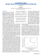
infrared (NIR) wavelength range (600 nm-1300 nm), often referred to as the tissue optical window, available for deep tissue diagnostics. A representa- tive plot of effective attenuation coeffi- cient2 in breast tissue as a function of optical wavelength is given in Fig. 1. Note the minimum (approximately 1.5 cm-1) in the 750-800 nm wavelength range, yielding a mean penetration depth of 0.66 cm. The optical fluence rate as a function of depth is shown in the inset of Fig. 1. Although the field decays quite rapidly (e.g., two orders of magnitude at a depth of 3 cm), diffuse
light has been successfully exploited for deep tissue charac- terization and imaging.
A variety of optical imaging techniques have been devel- oped for high resolution imaging in biological media. Excellent near-surface spatial resolution can be achieved using confocal optical microscopy and optical coherence tomography, but in both cases the image is formed using bal- listic (un-scattered) or quasi-ballistic light and thus is limited to depths3 of 1-2 mm. A pure optical imaging technique called diffuse optical tomography (DOT) has emerged as a powerful imaging modality that has been shown to be suit-
4
ablefordeeptissueimaging. Inthefrequencydomainimple-
mentation of this technique, intensity modulated laser light illuminates the media and an array of optical detectors meas-
Fig. 1. Effective attenuation coefficient of human breast tissue as a function of opti- cal wavelength. The inset shows the decay of the optical field as a function of depth in the 700-800 nm wavelength range.
Illuminating Sound 17
