
Page 21 - Summer 2007
P. 21
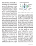
  Fig. 3. Acousto-optic imaging system using the charge-coupled device-based paral- lel speckle modulation processing approach.
 speckle. A key challenge in acousto-optic imaging is to understand how to detect the phase shift imparted on the modulated light given that the light levels, after passing through an optically diffuse media, are extremely low and come with a strong background of un-modulated light. If the aperture of an optical detector is restricted to detecting a sin- gle speckle, then one is left with the detection of a relatively small intensity modulation on an exceedingly weak signal. If the aperture is increased to receive light from multiple speck- le, the overall intensity of modulated light is increased, but the modulation depth is reduced owing to the random phase of the intensity fluctuations from speckle to speckle.
The idea of using ultrasound to localize or “tag” diffuse
light was first documented twenty years ago in a patent filed
by Dolfi and Micheron.7 The early 1990’s brought the first
experimental results demonstrating that the ultrasound
interaction with light in optically scattering media produces
a small, but measurable, intensity modulation in the scattered
field.8 During the mid 1990’s, researchers began using the
acousto-optic effect in order to image objects buried in tur-
9,10
Throughout this period, the majority of experi- ments were performed using a focused, continuous wave ultrasound beam to pump the acousto-optic interaction. The signal was detected using a single element optical detector that was fixed in space and apertured to sample a single speckle (or a small number of speckles) in the optical field. Images of absorbing objects were created by physically scan- ning the acoustic beam and measuring the intensity modula- tion of the optical field at each position. Continuous wave ultrasound allows for narrow-band detection techniques, and thus offers substantial improvements in signal-to-noise ratio over systems using pulsed ultrasound. However, since the interaction between light and sound occurs throughout the volume of the ultrasound field, this approach offers limited resolution along the ultrasound axis. To achieve axial resolu- tion from continuous wave exposures, a technique was intro- duced in which a single optical detector is used and the con- tinuous wave ultrasound source is rapidly chirped over a time frame comparable to the ultrasound propagation time
11
bid media.
In this way, a specific fre- quency is assigned to each location along the ultrasonic axis. A 1-D axial scan could then be produced from the time- dependent frequency-domain information of the ultra- sound-modulated signals. While acousto-optic imaging sys- tems using single optical detectors have been explored for a number of applications,12 the extremely low light levels asso- ciated with these systems are not conducive to deep tissue
sensing and imaging.
A breakthrough in the detection of ultrasound modulat-
ed signals came in 1999 with the development of a parallel
13
through the region of interest.
A schematic of the experimental setup is given in Fig. 3. In this approach, the single optical detector is replaced by a charge-coupled device (CCD) array, with the CCD camera positioned such that one speckle grain falls on each pixel. The main technical hurdle associated with this approach is that the speckle intensity modulation is in the MHz range, well beyond the frame rate of a CCD camera. To overcome this, the authors employ a
speckle modulation processing system.
 parallel lock-in detection scheme using synchronous excita- tion, rather than the more conventional synchronous detec- tion approach. The ultrasonic field and laser illumination source are both modulated at the same frequency (2.2MHz), with a fixed relative phase delay. The CCD camera acts as a low-pass filter and records the mean intensity of the field over the measurement time. Four images are recorded, each with the modulation of the laser source delayed by an additional one-quarter period with respect to the ultrasonic source. A linear combination of these images allows one to extract the amplitude and phase of the intensity modulation at each pixel. The amplitudes of the intensity modulations at each pixel are then summed to give the acousto-optic response. This allows for the coherent summation of signals over all detectors in the array and yields a signal to noise ratio improvement on the order of N1/2 over single detector schemes, where N is the number of pixels on the CCD. This CCD based approach has seen widespread use in acousto-optic imaging. Over the past several years a number of modifications have been proposed allowing, for example, improved sensitivity through hetero- dyne detection and operation with pulsed ultrasound for res-
14,15
interferometry technique has emerged that has sufficient sen- sitivity to detect acousto-optic signals generated by short-pulse
16
olution along the ultrasound axis.
More recently, a photorefractive crystal (PRC) based
ultrasound at clinically relevant acoustic exposure levels. Figure 4 illustrates the basic experimental setup used in the photorefractive crystal approach, where the sound source is the beam from a clinical diagnostic imager. The laser source is divided into a signal beam that illuminates the sample, and a reference beam that is sent directly to the photorefractive crys- tal. Scattered light from the sample is collected and mixed with the reference beam in the photorefractive crystal. The local index of refraction in the photorefractive crystal changes in response to the complex optical interference pattern set up in the crystal, and the reference beam diffracts off of this grating in a process called two-wave mixing. The crystal thus acts as a real time hologram, and the diffracted reference beam is an exact replica of the signal beam, less any high frequency (i.e., ultrasonic) modulation that exceeds the crystal response time. This diffracted reference beam is in-phase with the transmitted
Illuminating Sound 19
