
Page 20 - Volume 8, Issue 4 - Winter 2012
P. 20
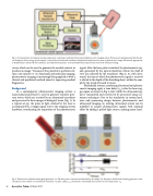
  Fig. 1. Instrumentation and major processing components comprising a combined ultrasound and photoacoustic imaging system. Photoacoustic signal, generated by the opti- cal absorption of laser energy by the sample, is received by an ultrasound transducer, beamformed and processed to form an photoacoustic image. Ultrasound, appropriate- ly beamformed, is emitted by the transducer, and reflected ultrasound is received, beamformed and processed to form the ultrasound image.
tracer, which can be used to generate the needed contrast to
14
In a contemporary photoacoustic imaging system, a nanosecond pulsed laser is used to generate transient pres- sure waves, which are received by an ultrasound transducer and processed to form images of biological tissue (Fig. 1). In a typical set-up, the pulse of light emitted by the laser is accompanied by a trigger signal sent to the imaging system hardware, coordinating the acquisition of the photoacoustic
signal. After the laser pulse is emitted, the photoacoustic sig- nals generated by the optical absorbers within the field of view are collected by the transducer (Fig 2). As with ultra- sound, the time at which the photoacoustic signal is received is related to the depth of the absorbing object within the sam- ple by the speed of sound in tissue.
Multimodal systems, combining ultrasound and photoa- coustic imaging, apply a time delay (τPA) after the laser trig- ger signal, as shown in Fig. 2, after which the ultrasound sig- nal is transmitted and received to be processed using cus- tomary techniques. Due to the similarities in system hard- ware and processing design between photoacoustic and ultrasound imaging, an existing ultrasound system can be modified to acquire photoacoustic signals with nominal effort by adding a pulsed light source, making minor hard-
Because of the potential to perform real- time, non-invasive in vivo functional and molecular imaging, photoacoustic imaging is increasingly being applied as both a clinical and preclinical method aimed at improving medical
diagnostics.
Background
produce an image.
 Fig. 2. Photoacoustic and ultrasound signal generation. (a) The laser pulse is scattered and absorbed by the sample. (b) Absorption which leads to heating generates a tran- sient pressure wave which is received by the transducer. (c) After a delay, τPA, ultrasound is transmitted and received over the time period 2· τPA.
16 Acoustics Today, October 2012
