
Page 21 - Volume 8, Issue 4 - Winter 2012
P. 21
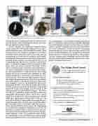
Fig. 3. Commercially available preclinical photoacoustic imaging systems.
ware modifications to appropriately time the transmitted and received signals, and by modifying the software user inter- face and signal processing methods.
Several companies are marketing integrated photoa- coustic systems for small animal imaging, shown in Fig. 3, and it is likely that more will soon enter the market. These systems fall into two categories—designed to use either a translatable linear ultrasound scanner with delay-and-sum beamforming, or a fixed array of transducers with comput- ed tomography image reconstruction. Linear photoacoustic scanning systems produce a two-dimensional (2D) view of a single projection, and are then scanned in a third dimen- sion – a volumetric imaging method analogous to com- monly used non-invasive clinical ultr asound systems, where a transducer array is manually scanned along the tis- sue surface. Tomographic systems use an array of transduc- ers, often positioned in a ring around the object being imaged, and then use reconstruction algorithms to repro- duce the imaged slice. Visual Sonics Inc. markets the Vevo LAZR (Fig. 3a), which uses their high resolution small ani- mal ultrasound imaging platform (Vevo 2100) in combina- tion with a tunable nanosecond pulsed laser. When whole body imaging is a priority, the multispectral optoacoustic tomography (MSOT) system (Fig. 3b), produced by iThera Medical, guides the laser beam to illuminate a ring of light around the animal being imaged, and then moves in the
15
choice of a scanning or tomographic system depends great- ly upon the application. The user must consider the achiev- able resolution, which is dependent upon the center fre- quency and bandwidth of the transducer, the beamforming or reconstruction algorithms implemented, and also the achievable sensitivity, which is influenced by the light flu- ence and distribution delivered within the tissue. An ultra- sound scanning system will generally be most flexible, accommodating a variety of animal types, and potentially providing multiple transducer arrays optimized for differ-
ent scanning depths. A photoacoustic imaging system based upon an ultrasound scanner platform provides additional functional imaging techniques (such as ultrasound Doppler mode) to provide anatomical and functional information. In contrast, tomographic systems are capable of acquiring volumetric data in a shorter period of time, and have less noise in the form of photoacoustic speckle due to the spatial distribution of the transducer ring. However, clinical appli- cations of a photoacoustic tomography system will be limit- ed, with a notable exception being the photoacoustic tomo-
graphic imaging of breast tissue.
16
The Walter Munk Award
Awarded in Recognition of Distinguished Research in Oceanography Related to Sound and the Sea
Call for Nominations
The Walter Munk Aw ard is s g gr ante d j o intly by
The Oceanography Society, the Office of Naval Research, and the Office of the Oceanographer of the Navy.
Recipients are selected based on their:
� ���������� �������� �������������� �� ��� ������������� �� �����-
cal ocean processes related to sound in the sea
� ���������� �������� �������������� �� ��� ����������� �� ��������
methods to that understanding
� ����������� ������� ���� ��������� �������� �� ����� ������� ���
instrum ent ati o n co ntrib utin g to o t th e ab ov e
For more information and nomination instructions, please visit: www.tos.org/awards_honors/munk_award.html
Nomination deadline is March 31, 2013.
THE OCEANOGRAPHY SOCIETY
The Oceanography Society, P.O. Box 1931, Rockville, MD 20849-1931, USA Telephone: 301/251-7708, Fax: 301/251-7709; E-mail: info@tos.org; Web site: www.tos.org
The Nexus 128 (Fig. 3c), produced by Endra, provides an alternative tomographic photoacoustic system. All of these systems consist of a tunable laser source providing near infrared (NIR) light approximately between 680 to 970 nm. The
third dimension to acquire volumetric images.
Photoacoustic Imaging for Medical Diagnostics 17
