
Page 22 - Volume 8, Issue 4 - Winter 2012
P. 22
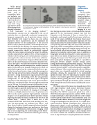
  Fig. 4. Transmission electron microscopy images of plasmonic metallic nanoparticles which have been used for in vivo photoacoustic imaging. (a) Gold nanospheres; (b) Silver nanoplates; (c) Gold nanorods coated with silica. Scale bars = 100 nm.
 While optical
absorbers naturally
found within the
body, such as
hemoglobin, lipids,
and melanin, can
be used to generate
photoacoustic
images, the use of
nano-sized imag-
ing contrast agents
allowsphotoacoustic
imaging to become
a truly “molecular” in vivo imaging method.17,18
Photoacoustic contrast can be enhanced by the use of
molecular dyes or chemically synthesized nanoparticles.
Inorganic nanoparticles are of particular interest, due to
their flexibility with respect to shape, size, and surface
chemistry. These characteristics can be optimized for
enhanced delivery of the nanoparticles to the tissue of
interest, such as cancerous tumors.19 If the nanoparticle sur-
face is modified by the addition of a targeting moiety, these
contrast agents become molecular imaging agents, since the
targeted nanoparticles should be preferentially retained
within the tissue region expressing the target. In addition,
metallic nanoparticles of gold or silver, several examples of
which are shown in Fig. 4, have surface plasmon resonance,
meaning that the valence electrons of the atoms collective-
ly oscillate at a characteristic frequency. When the incident
light is at the same frequency as the surface plasmon reso-
nance, that light is very efficiently absorbed and converted
into heat, making metallic nanoparticles excellent contrast
agents for photoacoustic imaging. Nanoparticles which are
molecularly targeted can be customized for delivery to par-
ticular tissues based on size, shape, and surface properties,
while the high optical absorption of metallic nanoparticles
generates a strong photoacoustic signal and provides high
contrast with the surrounding tissue. By using metallic
nanoparticles, we can also tune their optical absorption
properties to take advantage of the “tissue optical window”.
Within this wavelength region, between approximately 600
nm to 1300 nm, the native tissue optical absorption is rela-
tively low, and therefore light can penetrate deeper into tis-
sue, allowing for acquisition of photoacoustic images at sig-
nificantly greater tissue depths. By imaging at wavelengths
which are not highly absorbed in tissues, the photoacoustic
effect can be used to generate high resolution molecular
images in vivo at significant tissue depth. Questions remain
regarding the safety of metallic nanoparticles for use in the
20,21
of nanoparticles.
Diagnostic photoacoustic imaging
In our studies, anatomical, func- tional, and molecu- lar information was acquired using a combination of ultrasound and multiwavelength photoacoustic imaging of in vivo
mice bearing cancerous tumors. All methods follow protocols approved by the Institutional Animal Care and Use Committee at the University of Texas at Austin. First, we developed a small animal model of breast cancer which con- sisted of two tumors established from human breast cancer cell lines with differential cell biomarker expression. For this small animal model, we initiated tumors within the mamma- ry fat pad using injections of BT-474 cancer cells, which over- express the cellular receptor HER2, and MDA-MB-231 cancer cells, which over-express αvβ3 integrin, present on the cell sur- face of epithelial cells of neovasculature. Gold nanorods were chosen as the molecular contrast agent since, in addition to being highly optically absorbing due to surface plasmon reso- nance effects, their optical absorption spectra can be tuned by
23
We coated the nanorods with amorphous silica, which improves the gold nanorod thermal stability25 and the photoacoustic signal gen-
Indeed, there is much that is still unknown about both their short-term and long-term tox- icity, but many groups are studying these effects,21 and three clinical studies of gold nanoparticle-based method- ologies for treatment of cancer are underway or have been completed.22 Additionally, photoacoustic imaging could play a role in improving the understanding of the biodis- tribution and clearance mechanisms affecting the toxicity
system was used to deliver between 10-20 mJ/cm
human body.
18 Acoustics Today, October 2012
By tuning nanorods to have dif- ferent peak optical absorption wavelengths, it is possible to distinguish between multiple nanorod contrast agents
24
Silica-coated nanorods with two different
changing their aspect ratio.
through multiwavelength photoacoustic imaging.
eration efficiency.
26
aspect ratios were chemically modified to attach targeting
antibodies, allowing the nanoparticle to be preferentially
uptaken by tumor cells over-expressing the targeted cellular
24
The tumor region of the mouse was imaged using a Vevo 2100 high frequency small animal ultrasound scanner (VisualSonics Inc.), integrated with a SpectraPhysics QuantaRay Pro Nd:YAG nanosecond pulsed laser with a GWU PremiScan optical parametric oscillator (OPO) to tune the light wavelength. A fiber optic bundle was used to deliver the laser light on either side of a linear transducer array. The
2
The contrast agents were injected into the blood- stream of a mouse growing the tumors. The injected contrast agents distributed through the circulatory system, and infil- trated through the tissues of the mouse, including the cancer- ous tumors.
receptor.
of light over a range of wavelengths from 680-930 nm. A 21-MHz ultra- sound transducer (MS250, VisualSonics Inc.) collected the photoacoustic signals at each wavelength, followed by the transmission of ultrasound and the receiving of the reflected ultrasound by the same transducer, resulting in co-registered ultrasonic and photoacoustic images. A linear stepper motor was used to translate the transducer and fiber bundle con- struct, in steps of 150 μm, to acquire photoacoustic and ultra-
