
Page 15 - Jan2013
P. 15
MICROBUBBLES AS ULTRASOUND CONTRAST AGENTS
Thomas J. Matula
Applied Physics Laboratory, University of Washington Seattle, Washington 98105
and
Hong Chen
Department of Biomedical Engineering, Columbia University New York, New York 10027
Introduction
The future of healthcare is bubbles.
That may be an overstatement, but
micron-sized bubbles (called
microbubbles) play an important role in diagnostic imaging. Current research is exploring how microbubbles can be used for molecular imaging and targeted drug delivery. The bubbles act as very good ultrasound scatterers, and because they oscil- late upon ultrasound exposure, they can also do (therapeutic) work on the surrounding tissue (e.g., breaking blood clots or opening up the blood-brain barrier). Current research with microbubbles has focused on two main areas—developing new ultrasound pulse sequences to improve the contrast/noise ratio, and developing specialized microbubbles for molecular imag- ing and therapy. This article discusses aspects of microbubbles and their dynamics in actual blood vessels.
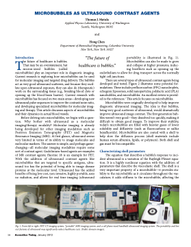
Before delving into microbubbles, we begin with a ques- tion. Why bother with ultrasound as a molecular imaging/therapy modality? Molecular imaging is already being developed for other imaging modalities such as Positron Emission Tomography (PET) and Magnetic Resonance Imaging (MRI). At first glance, ultrasound seems very limited in terms of its ability to image molecules and molecular markers. The answer is simple, and perhaps game- changing—all molecular imaging modalities require some sort of contrast agent. Gadolinium-based agents are examples of MR contrast agents; fluorine 18 is an example for PET. With the addition of ultrasound contrast agents like microbubbles that are targeted to specific antigens, ultra- sound too has the potential of being able to image disease proteins at the molecular level. Ultrasound has the added benefits of being low cost, non-invasive, highly portable, uses no radiation, and allows for real time imaging (ultrasound
portability is illustrated in Fig. 1). Microbubbles can also be made to grow and collapse at higher pressures, induc- ing bioeffects such as opening up the
endothelium to allow for drug transport across the normally tight cell junctions.
There are several types of ultrasound contrast agents being developed and tested. Figure 2 illustrates some potential for- mulations. These include perfluorocarbon (PFC) nanodroplets, echogenic liposomes, solid nanoparticles, polylactic acid (PLA) nanobubbles, and microbubbles. An excellent review is provid- ed in the references.1 This article focuses on microbubbles.
Microbubbles were originally developed to help improve diagnostic ultrasound imaging. The idea is that bubbles, being very good scatterers of ultrasound, would dramatically improve ultrasound image contrast. The first generation bub- bles weren’t very good—they dissolved too quickly, making it difficult to obtain good images. To improve their stability, today’s microbubbles are filled with heavier gases of lower solubility and diffusivity (such as fluorocarbons or sulfur hexafluoride). Microbubbles are also coated with a shell to help slow the diffusion process (shell materials include human serum albumin, lipids, or polymers). Both shell and gas must be biocompatible.
Characterizing shell parameters
The equation that describes a bubble’s response to inci- dent ultrasound is a variation of the Rayleigh-Plesset equa- tion. It is a highly nonlinear equation with the addition of parameters that describe the viscoelastic shell. The shell is a very important property of a microbubble—it provides sta- bility to the microbubble as it circulates throughout the vas- culature; it adds stiffness to the microbubble, affecting the
“The future of healthcare is bubbles.”
Fig. 1. From left to right: PET-CT imaging system, “portable” MRI imaging system, and a cell phone-sized handheld ultrasound imaging system. The portability and low cost features of ultrasound may significantly reduce healthcare costs. (Public domain images).
14 Acoustics Today, January 2013
