
Page 43 - 2016Summer
P. 43
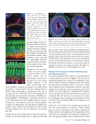
Figure 2. A: Mammalian co- chlea with hair cells (green), nerves (red) and spiral ganglion neurons (yellow/orange). B: In- ner hair cells and stereocilia (green), with nuclei (blue) and nerve fibers of neurons that transmit information to the brain (red; McLean et al., 2009). C: Top of the three rows of outer hair cells (green dots at top) and the tubular single row of inner hair cells innervated by neural process (red/orange).
transmits highly selective in- formation about the frequen- cy, timing, and intensity of sounds to the brain. Support- ing cells are nonsensory cells that neighbor and isolate hair cells from one another. These nonsensory cells work with the surrounding structures to provide physical and mo- lecular support to this elabo- rate sensory epithelium.
Hearing loss can result from a failure of acoustic signals to reach the inner ear (con- ductive hearing loss) or from damage to any part of the inner ear or the central auditory pathways in the brain (sensorineural hear-
ing loss[SNHL]). Conductive hearing loss is usually treated by medical or surgical means. The most common form of SNHL results from damage or dysfunction of hair cells in the organ of Corti. When hair cells in the mammalian co- chlea die, they do not regenerate; this form of SNHL is per- manent (Figure 3). If hearing loss is moderate, patients can be fit with hearing aids, which amplify sounds to enhance hearing. If it is severe, patients can receive cochlear implants to bypass the injured hair cells and directly stimulate the auditory nerve. Neither form of treatment restores normal hearing or addresses the cause of hearing loss, the missing hair cells.
Around 30 years ago, the discovery that hair cells regenerate in birds raised the possibility that we could someday find
Figure 3. Left: Surface view of a healthy cochlea with hair cells (green), neural processes (red), and nuclei (blue).Right:A damaged cochlea that no longer contains hair cells but has preserved neural processes and nuclei. White arrows: Organ of Corti boundaries.
away to replace hair cells in mammals, including humans. Since that time, many advances in our understanding of hair cell regeneration in birds, fishes, and mammals have been achieved. This article reviews the current state of research in the field of hair cell regeneration. Due to space limitations, we have removed all but the most essential citations. For fur- ther details and relevant citations, we encourage readers to examine the many review papers related to this field (e.g., Warchol, 2011; Groves et al., 2013; Rubel et al., 2013).
Cellular Processes of Hair Cell Damage and Regeneration
The sensory epithelium of the cochlea is a cytoarchitectural- ly elegant and delicate structure (Figure 1). The hair cells are commonly damaged by a variety of environmental events, some of which are known, including acoustic overstimula- tion from loud or prolonged noise or concussive stimuli. Several different types of medications kill hair cells when administered at high doses or for prolonged periods. These include, but are not limited to, aminoglycoside antibiotics such as gentamicin and heavy metal anticancer drugs such as cisplatin. Hair cells also die as we age; in most cases, this is due to unknown causes. Finally, genetic mutations exist that cause hair cells to die during embryonic development or at later stages of life.
Until 1985, it was believed that regeneration of inner ear hair cells was not possible in vertebrates. While studying processes of hair cell damage in the chicken auditory epi- thelium, however, investigators noted a reappearance of hair cells in the area of damage. The immature morphology of these cells appeared similar to that of embryonic hair cells in
    Summer 2016 | Acoustics Today | 41
