
Page 44 - 2016Summer
P. 44
Regeneration of Auditory Hair Cells
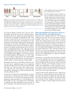
Figure 4. The undamaged auditory epithelium of the bird contains hair cells (HC; red) interdigitated with supporting cells (SC; white). On damage, hair cells are removed from the epithelium and supporting cells are triggered to regenerate hair cells. Nonmitotic re- generation allows a supporting cell to change its shape and genetic profile to that of a hair cell. Mitotic regeneration requires a supporting cell to divide and differentiate into two daughter cells, a hair cell and a supporting cell.
newly formed cells become replacement hair cells (Oesterle et al., 2003).
The big challenge facing researchers to- day is to determine why hair cells are not readily regenerated in mammals. Regen- eration could fail in the adult cochlea be- cause the hearing organ loses the popula- tion of progenitor cells capable of forming new hair cells during development. Alterna- tively, cells with the potential to replace hair cells may exist in the cochlea but are unable to respond to damage due to active inhibi- tion or lack of stimulatory signals.
the cochlea of chickens (Cotanche, 1987; Cruz et al., 1987). During this same period, it was also discovered that regener- ation of hair cells occurs readily in the vestibular portions of the avian inner ear (Jørgensen and Mathiesen, 1988). Soon, researchers learned that the hair cells of the inner ear and lateral line system of fish, frogs, and salamanders also read- ily regenerate after damage, which led to the conclusion that regeneration occurs in hair cell epithelia of all vertebrates except mammals. Further analysis revealed that the support- ing cells that normally surround the hair cells are the source of these newly differentiating hair cells. Supporting cells may either mitotically divide to achieve hair cell differentiation or phenotypically convert to a hair cell in a process called di- rect transdifferentiation (Figure 4) (Corwin and Cotanche, 1988; Ryals and Rubel, 1988; Roberson et al., 1996). With these two methods of replacing hair cells, nonmammalian vertebrates provide valuable models to study these processes and their ability to restore hearing after sustained SNHL.
In mammals, the situation is quite different. When hair cells die in the mature mammalian organ of Corti, supporting cells fill in the gaps where hair cells were located to form permanent scars, and no new hair cells are formed. More- over, supporting cells neither divide nor convert into hair cells after hair cell damage (e.g., Roberson and Rubel, 1994; Chardin and Romand, 1995).
In contrast to the organ of Corti, adult mammals can sponta- neously replace a small number of hair cells in the vestibular organs of the inner ear. New hair cells are largely formed by nonmitotic regeneration (Forge et al., 1998; Kawamoto et al., 2009; Golub et al., 2012). There appears to be a small degree of supporting cell division triggered in response to hair cell loss (Li and Forge, 1997; Kuntz and Oesterle, 1998), but no
Stimulating Native Progenitors to Form New Hair Cells in the Adult Cochlea Researchers have examined whether the cells capable of forming new hair cells still exist in the cochlea of mature mammals. Many tissues in our body undergo continual renewal. One common feature of these tissues is that they contain stem cells that divide and form new specialized cells throughout life. Several lines of evidence show that the co- chlea and vestibular organs possess stemlike progenitors to hair cells during early development but lose them as the or- gans mature (Oshima et al., 2007). Consistent with this, new hair cells can be formed by supporting cells from the organ of Corti of neonatal mammals (White et al., 2006; Cox et al., 2014), but not in adult mammals (e.g., Roberson and Rubel, 1994; Forge et al., 1998).
Investigators are using three general strategies to identify ways to trick supporting cells in the mature mammalian in- ner ear to regenerate hair cells. First, we are finding clues in cochlear development. Hair cells in the organ of Corti form during the embryonic period through a complex series of cellular steps controlled by a cascade of molecular interac- tions. Some researchers have postulated that, before any cell in the mature cochlea can form a new hair cell, it will need to relive these same stages of development.
Second, we look to other regenerative tissues. Many tissues in the body are continuously replaced under normal condi- tions and/or after damage, including cells in the skin, intes- tine, and some regions of the brain. We reason that many of the molecular cascades leading to regeneration in these other tissues could be co-opted to trigger regeneration in the cochlea.
42 | Acoustics Today | Summer 2016
