
Page 45 - 2016Summer
P. 45
Third, using the new tools of molecular genetics, we can directly query the molecular cascades that are activated in the sensory epithelia of nonmammalian vertebrates that do regenerate hair cells, such as birds and fishes. In the section below, we describe several genes and signaling pathways that met one or more of these criteria and were evaluated for their capacity to stimulate hair cell regeneration in mam- mals. These analyses revealed signaling molecules that are important for facilitating regeneration.
Forced Atoh1 Expression: Pushing Mature Supporting Cells to Transdifferentiate Into Hair Cells
A proneural transcription factor named atonal homolog 1 (Atoh1) is a potential therapeutic agent for promoting hair cell regeneration. Atoh1 helps to direct the generation of hair cell-specific proteins that give the hair cell its morpho- logical and physiological identity (Cai et al., 2015). When the gene encoding Atoh1 is deleted, hair cells in the organ of Corti do not form (Bermingham et al., 1999). Thus, Atoh1 is a very powerful activator of hair cell features and could trig- ger cells to transdifferentiate into hair cells.
In tissues that regenerate hair cells, Atoh1 expression is activated in supporting cells shortly after hair cell damage (Cafaro et al., 2007; Wang et al., 2010; Lin et al., 2011). In cultured auditory organs from chickens, forced expression of Atoh1 influences supporting cells to form new hair cells by promoting division and direct transdifferentiation (Lewis et al., 2012). In rodents, forced expression of Atoh1 by viral injection into the organ of Corti or nearby regions of de- veloping mice forces more cells to differentiate as hair cells (Zheng and Gao, 2000; Gubbels et al., 2008). These findings suggested Atoh1 misexpression might be sufficient to trig- ger supporting cells to transdifferentiate into hair cells af- ter damage in the cochlea of adult mammals. Indeed, some studies suggest that Atoh1 may drive production of new hair cells in auditory (Izumikawa et al., 2005) and vestibular (Schlecker et al., 2011) organs, which might result in small improvements in hearing and balance function.
However, recent studies are less encouraging. Misexpres- sion of Atoh1 in pillar and Deiters’ cells, two supporting cell subtypes (Figure 1), in the mature mouse cochlea stimulates early stages of transdifferentiation into hair cells, but this process is not completed and many “forced” cells die (Liu et al., 2012). Indeed, Atkinson et al. (2015) noted no sig- nificant improvement in hearing after virally induced Atoh1 misexpression in the organs of Corti of guinea pigs. Hence, an important current challenge is to determine what factors
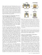
Figure 5. Expression patterns of the Notch receptor and Atoh1 tran- scription factor in supporting cells (yellow) and hair cells (blue) un- der normal, damaged, and regenerating conditions. In supporting cells in undamaged epithelia, there is high Notch receptor activity and the Notch intracellular domain (Notch ICD) travels to the nucleus, inhibiting Atoh1 expression. In supporting cells after hair cell dam- age, Notch receptor activity is reduced, Notch ICD remains at the membrane, and Atoh1 levels increase, driving the supporting cell to transdifferentiate into a hair cell. Once the new hair cell matures, Notch activity is increased again and Atoh1 transcription is reduced to normal levels.
limit the ability of Atoh to drive hair cell regeneration in the mature cochlea. Currently, a human clinical trial testing the ability of viral infection of Atoh1 to improve hearing is un- derway. Results are not available at this time.
Suppression of Notch Signaling: Can This Enhance Proregenerative Effects of Atoh1?
As discussed above, it is evident that, although Atoh1 mis- expression reliably promotes supporting cells and other cells around the organ of Corti to become hair cells in neonatal mammals, unidentified factors appear to hinder the effects of Atoh1 in the mature organ of Corti. One likely suspect is the factor is signaling through the Notch receptor (Lewis, 1998).
Notch is a receptor on the surface of cells that is activated by molecules on adjacent cells (Figure 5). Notch has many functions in a variety of cells, but its most pertinent role with respect to hair cell regeneration is the inhibition of hair cell formation. During development, Notch ligands are expressed in young hair cells and influence surround- ing supporting cells to maintain their identity rather than differentiate into hair cells (reviewed in Kelley, 2006). Notch signaling executes this function, at least in part, by block- ing Atoh1 synthesis (Lanford et al., 2000). In the developing
Summer 2016 | Acoustics Today | 43
