
Page 39 - 2018Fall
P. 39
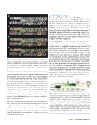
 Figure 2. Visual analogy of parsing an auditory scene. a: Different sources overlapping each other, making them hard to separate. b: Acoustical features, here depicted in different colors, help separat- ing auditory objects. c: Degraded signals can make scene analysis more difficult. d: Attention can be maintained or switched across two groups of texts.
tures are degraded such as in a highly reverberant concert hall, auditory scene analysis is even more difficult (Figure 2c). Finally, communication in social settings often involves selectively maintaining attention to a particular auditory ob- ject (e.g., your colleague giving constructive criticism on the paper you just presented) and switching attention to other sounds in the environment (e.g., the waiter just passed by asking whether you care for a canapé). This flexibility of at- tending one sound source and then switching to another at the next instant (e.g., switching attention between two groups of texts in Figure 2d as a visual analogy) is the hall- mark of active listening.
How does the brain accomplish this active listening task? Here, I describe our current understanding of how the brain analyzes an auditory scene and forms auditory objects as well as how neural activity in the brain is modulated when we selectively attend to one sound in a mixture. But first, I briefly describe the neuroimaging techniques we use to study the listening brain.
Imaging Techniques
Functional Magnetic Resonance Imaging
Functional magnetic resonance imaging (fMRI) is a widely used noninvasive neuroimaging technique. When part of the brain works harder, its oxygen consumption increases; fMRI measures this blood oxygenation level-dependent (BOLD) signal. Even though the BOLD signal is only a proxy for neural activity, there is good correspondence of local field potential reflecting the underlying neural activ- ity and the BOLD response (Logothetis, 2008). This neuro- imaging technique has excellent spatial resolution but poor temporal resolution (Figure 3).
Acoustically, fMRI as a neuroimaging technique presents a unique challenge, especially in studies involving auditory at- tention. Noise in the fMRI environment can reach 110-dB sound pressure level (SPL), approximately as loud as a chain- saw. Hearing protection is used to mitigate noise exposure, but this scanner noise is an unavoidable component of the auditory scene that a subject hears during an fMRI study and can directly affect auditory experimentations (Cusack, 2005). An alternative strategy is to take brain images not continuous- ly but sparsely in time and present an auditory stimulus dur- ing the silent periods between imaging acquisitions (Hall et al., 1999). However, this approach lowers the signal-to-noise ratio of the image, and the corresponding sporadic scanner noise can potentially be more distracting than having scanner noise present continuously. These factors illustrate the chal- lenges experimenters face when designing psychoacoustic ex- periments in an fMRI environment.
Figure 3. Trade-offs across neuroimaging modalities in terms of spa- tial resolution, temporal resolution, and brain coverage. Each neuro- imaging technique has its own strengths and weaknesses. Functional magnetic resonance imaging (fMRI) has millimeter resolution but temporal resolution is worse than with other techniques. Magneto- encephalography (MEG) and electroencephalography (EEG; M/EEG when combined) have millisecond temporal resolution but poorer spatial resolution. Electrocorticography (ECoG) has both great spa- tial and temporal resolution but can only record part of the brain, whereas the other techniques can cover the whole brain.
   Fall 2017 | Acoustics Today | 37
