
Page 55 - 2017Winter
P. 55
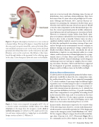
 Figure 1. Diagram showing key structures in a cross section of a human kidney. The top of the figure is toward the head and the artery and vein point toward the center of the body. Filtra- tion and fluid extraction occurs in the renal cortex and renal pyramids from blood that enters via the renal artery and, after filtration, exits via the renal vein. The filtrate is then trans- ferred through tubules in the pyramids to the collecting space in the calyx. Urine then passes down the ureter to the bladder.
Figure 2. Center microcomputed tomography (μCT) slice of a calcium oxalate monohydrate (COM) kidney stone. Gray structures in the image show the inorganic crystals; dark or radiolucent regions are indicative of the organic protein ma- trix and may contain pockets or trapped gas or microbubbles. Image courtesy of James C. Williams, Jr.
nauts are at an increased risk of forming stones because of dehydration, stasis, and bone demineralization. There have been more than 32 stones observed postflight in US astro- nauts (Sibonga and Pietrzyk, 2017), and one Russian cos- monaut was preparing for emergency deorbit from space when his stone and the mission critical crisis passed. Should a stone become symptomatic on the International Space Sta- tion, the current operational protocol includes administra- tion of pharmaceuticals and emergency evacuation to Earth. However, as astronauts venture farther from Earth, emer- gency evacuation is unfeasible because transit increases from hours to days, weeks, or months. Urinary stones are also a significant military medical concern because they result in lost duty days and medical evacuations (average 60 per year) and are brought on by environmental stressors common to military training and operational settings (Armed Forces Health Surveillance Branch, 2011). Specifically, the risk of dehydration is high for an unacclimated soldier in a warm climate, and the mean interval from deployment to a symp- tomatic stone is 93 days (Evans and Costabile, 2005). For both NASA and DoD, clinical technologies used to diagnose and manage stones are too large or complex, thus necessitat- ing evacuation. Therefore, NASA and DoD have supported the development of portable, noninvasive technologies to diagnose and treat kidney stones in the field or in flight.
Kidney Stone Detection
To advise patients on management options for kidney stones, physicians would like to know the size, composition, num- ber, and location of the stones. X-ray computed tomography (CT), which combines X-ray images taken at different angles to produce cross-sectional images or slices is considered the gold standard for diagnosing stones. CT images of stones re- quire little interpretation for physicians to (1) identify the stone position and kidney structures, (2) predict something about stone composition or susceptibility to breakage based on the intensity of the stone, and (3) accurately size the stone (Figure 3a). However, CT also exposes patients to ionizing radiation that increases the risk of developing cancer and limits its use for routine monitoring (Fwu et al., 2013).
In most studies, ultrasound is reported as less sensitive than CT for detecting stones; however, a recent study demonstrat- ed ultrasound as statistically equivalent to CT for diagnosing stones in the emergency room (Smith-Bindman et al., 2014). Yet few surgeons will go to surgery based on ultrasound im- ages alone (Ganesan et al., 2017). Ultrasound (Figure 3b) is less commonly used to detect kidney stones in the United
   Winter 2017 | Acoustics Today | 53
