
Page 57 - 2017Winter
P. 57
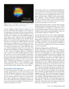
 Figure 4. The color Doppler ultrasound twinkling artifact highlights kidney stones with a mosaic of colors.
To make twinkling a reliable detection technique, it is im- portant to understand why twinkling occurs. Machine jit- ter, radiation force that moves the stone, and internal rever- berations within the stone have all been proposed to cause twinkling. The oscillation of cavitation bubbles trapped in stone surface crevices could also cause the backscattered signal to differ between each pulse of the ensemble. This hy- pothesis was recently tested by exposing stones to elevated static pressure that compresses bubbles and drives them into solution because solubility increases with pressure in accor- dance with Henry’s law. In a water bath, twinkling on stones was found to be suppressed by elevated hydrostatic pres- sure. Similarly, improved wetting of the stone surface with alcohol to remove bubbles also suppressed twinkling (Lu et al., 2013). The spectral Doppler signatures of twinkling on stones resemble those from gas-filled ultrasound contrast agents (Bruce et al., 2016), and microbubbles entrenched on the stone surface have now been directly observed with a high-speed camera after enlarging the bubbles with a modi- fied lithotripter pulse (Simon et al., 2017). Future work in- cludes determining the distribution and contents of the surface cavitation bubbles as well as modeling the effect of elevated pressures on the resonance of crevice bubbles, which may help elucidate why only some stones twinkle.
Kidney Stone Management
The preferred management for kidney stones, when small enough, is observation to see if the stone will pass on its own. The likelihood of spontaneous stone passage depends on stone size: 80% of small stones less than 5 mm in diam- eter pass spontaneously, but less than 30% of stones greater than 6 mm in diameter pass spontaneously (Ueno et al., 1977). Some medications that are thought to facilitate spon- taneous stone passage are available, but their effectiveness is controversial (Somani et al., 2016).
If a stone does not pass or is accompanied by complications such as infection or obstruction, more proactive manage- ment options such as ureteroscopy, shock wave lithotripsy (SWL), and percutaneous nephrolithotomy (PCNL) are em- ployed. In ureteroscopy, a flexible ureteroscope with fiber- optic capabilities is inserted through the urethra, bladder, and ureter and into the kidney. Lasers inserted through the ureteroscope are then used to pulverize the stone to frag- ments that are passable or can be extracted.
Introduced in the 1980s (Chaussy et al., 1980), SWL is the only completely noninvasive treatment for kidney stones. In SWL, an electrohydraulic, electromagnetic, or piezoelectric source outside the patient generates shock waves that are focused on the kidney stone to shatter it into small fragments so they can pass through the urinary tract (Bailey et al., 2006).
Ureteroscopy and SWL are used in nearly equal propor- tion and account for over 90% of cases (Bach and Buchholz, 2011). PCNL is employed in rarer, more complex cases in- volving patients with many or very large stones and entails inserting a 1-cm-diameter tube into the kidney through the back. All procedures have associated injury to the kidney or ureter and risks of complications that research and patient selection can minimize.
Kidney Stone Fragmentation with Ultrasound
In SWL, shock waves act to break apart a stone through two dominant mechanisms: elastic waves and acoustic cavitation (Zhu et al., 2002; Sapozhnikov et al., 2007). Although elastic waves generate large dynamic tensile stresses within the stone body, producing fractures, acoustic cavitation bubbles in the fluid surrounding the stone grow and collapse against the surface, causing hydrodynamic jetting, surface erosion, and pitting. Researchers have sought to improve lithotripters by enhancing stress waves or cavitation. For instance, it has been suggested that a broader focus can achieve better shear wave production in a stone and enhance fractures (Eisenmenger, 2001). Others have proposed altering the waveform of the shock wave to better localize cavitation around the stone (So- kolov et al., 2001). However, these bubbles remain in the fluid after a shock and can rapidly multiply into a large cloud (Pish- chalnikov et al., 2006). The result is attenuation of subsequent shocks and shielding of the acoustic energy from reaching the stone, leading to diminished fragmentation (Paterson et al., 2002). This effect limits the rate at which physicians operate a lithotripter to 1-2 shocks per second (to give bubbles time to dissolve), with clinical results showing increased safety and effectiveness at slower rates (Pace et al., 2005).
 Winter 2017 | Acoustics Today | 55
