
Page 56 - 2017Winter
P. 56
Kidney Stones and Ultrasound
States because of the high availability of CT and the dependence of ultra- sound on the skills and interpretation of the user. In the ideal ultrasound image, kidney stones appear as the brightest object in the image, with a posterior acoustic shadow due to the high acoustic impedance of the stones compared with the surrounding soft tissue. However, the stones do not al- ways appear appreciably brighter than the surrounding tissues because the surface curvature or small size of a stone (relative to the ultrasound wave- length) do not necessarily scatter sound directly back to the transducer.
Another challenge in detecting stones with ultrasound is that commercial ultrasound scanners are optimized to detect subtle changes in soft tissue properties, resulting in poor contrast and resolution when imaging stones. Improvements can be made to optimize kidney stone detection. For exam- ple, ultrasound machines compress the voltage range of the received sig- nals to display the intensity of the backscattered signal in a limited num- ber of grayscale levels. Rather than map linearly, the voltage is mapped to intensity on a curve that, for most machines, accentuates the difference between the weak signals reflected from tissue and compresses all stron- ger reflections to the top few bins of brightness. Altering the compression curve to accentuate the strong stone reflections can make stones easier to detect but sacrifices sensitivity to differentiate soft tissues. Similarly, averaging images made from several angles smooths the image, which is desirable for tissues but washes out the strong contrast of the stone and its shadow. These improvements have already been shown to reduce stone size overestimation by 1.6 ± 1.0 mm (May et al., 2016a) compared with overestimates as high as 3.9 ± 1 mm with clinical ultrasound (Sternberg et al., 2016). Size accuracy was further improved, with a difference of only 0.8 ± 0.6 mm (compared with CT), by measuring the width of the posterior acoustic shadow rather than the width of the stone in the image (May et al., 2016a). The reason a stone itself appears large but the shadow does not is not well understood.
Another way to make a stone more conspicuous in an image is to use the Doppler ultrasound “twinkling artifact” (Rahmouni et al., 1996; Figure 4). In Doppler ultrasound, an ensemble of ultrasound pulses is transmitted and the variability in the backscattered pulses of that ensemble is evaluated through correlation to detect changes that are displayed as color overlaid on a grayscale image. Twinkling occurs when each pulse in the ensemble differs in a nondeterministic way, causing the ultrasound machine to high- light the stone with a mosaic of changing colors. If pulses differ determin- istically from a phase or time lag due to the target moving away from the probe as in blood flow, a solid color representing flow velocity appears on the screen (Kremkau, 2010). Whereas stones in grayscale ultrasound im- ages are on average no brighter than the brightest background tissue, the signal strength on a twinkling stone is on average 37 times stronger (Cu- nitz et al., 2017). Although twinkling can make stones very conspicuous, not all stones twinkle, and at times, modern machines show twinkling on image features that are not stones (Masch et al., 2016).
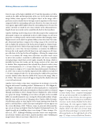
Figure 3. Imaging modalities commonly used to detect kidney stones: computed tomography (CT; a) and ultrasound (b) in two different hu- man subjects. In the CT slice, the stone appears brighter than the surrounding tissue and kidney structures can be clearly identified. In the ultra- sound image, the stone appears brighter than the surrounding tissue with a posterior acoustic shadow, which is the ideal case of stone imag- ing with ultrasound, Image is oriented with the ultrasound probe on the right-hand side of the page. CT image courtesy of Barbrina Dunmire.
54 | Acoustics Today | Winter 2017
