
Page 37 - Summer 2018
P. 37
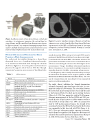
           Figure 1. a: Bones consist of two types of tissue: cortical and cancellous. As osteoporosis progresses, the cortical layer be- comes thinner and the cancellous bone becomes more porous. b: High-resolution X-ray computed tomography images show- ing low- and high-density specimens of cancellous bone excised from the femur (thigh bone) near the location shown in a.
Clinical Ultrasound Devices for Bone Fracture Risk Assessment
The earliest and best-validated design for a clinical bone ultrasound device uses a broadband ultrasound transduc- er to transmit an ultrasound beam through the calcaneus (heel bone; Figure 3a). A receiving ultrasound transducer is placed on the opposite side of the foot. With this “through- trWanesamri–ssiAonrt”icgleo4metry, measurements of broadband ultra-
Figure 2. Acoustic impedance images of human cortical bone transverse cross sections from the tibia (long bone of the lower leg) measured at 100 MHz. a: Healthy specimen; b: late-stage osteoporotic specimen showing dramatic thinning of cortical bone. See Raum et al., 2008.
sound attenuation (BUA) and speed of sound (SOS) may be performed. Both BUA and SOS have been shown to be high- ly correlated with calcaneal BMD. Attenuation in bone is far greater than attenuation in soft tissues so attenuation due to soft tissue surrounding the calcaneus has little impact on the measurements. Using a broadband ultrasound transducer allows attenuation to be measured at multiple frequencies (typically, about 300-700 kHz). Attenuation in bone increas- es with frequency and is usually characterized by the slope of a linear fit to attenuation versus frequency (BUA; see The Interaction of Ultrasound with Cancellous Bone). The SOS may be estimated from the time delay between transmitted and received ultrasound pulses.
The initial clinical validation for through-transmission calcaneal ultrasound came from large-scale trials. In a ret- rospective study of 4,698 women, the association between BUA at the heel and at existing fractures was nearly the same as that between DXA at the heel and at existing fractures (Glüer et al., 1996). In a prospective study of 5,662 women, ultrasonic measurements at the heel (BUA and SOS) pre- dicted the risk of hip fracture in elderly women nearly as well as DXA at the hip (Hans et al., 1996). In a prospective study of 6,189 postmenopausal women, the strength of the association between the BUA at the heel and at the fracture was comparable to that observed with DXA (Bauer et al., 1997). These and subsequent studies represent the founda- tion for formal recognition of the diagnostic effectiveness of calcaneal ultrasound by professional organizations (Krieg et al., 2008; US Preventive Services Task Force, 2011).
   Table 1.
AbAbrreevviaiatitoionnss
Term
     Definition
     AT
   Axial (along the axis of long bones) transmission
    BDA T
      Bidirectional axial transmission
  BMD
  Bone mineral density (in g/cm2 for projection methods like DXA or g/cm3 for volumetric methods like QCT)
    BUA
   Broadband ultrasound attenuation (in dB/MHz)
    DXA
   Dual-energy X-ray absorptiometry
    FAS
   First arrival signal
    FFGW
   Fundamental flexural guided wave
    HR
      High resolution
  HR-pQCT
  High-resolution peripheral quantitative computed tomography
    nBUA
   Normalized (to bone thickness) BUA (in dB/cmMHz)
    pDXA
   Peripheral dual-energy X-ray absorptiometry
    pQCT
   Peripheral (arms, legs, hands, feet) QCT
    QCT
   Quantitative computed tomography
    QUS
   Quantitative ultrasound
    SOS
   Speed of sound (in m/s)
    TOF
    Time of flight (in s)
 Summer 2018 | Acoustics Today | 35
