
Page 39 - Summer 2018
P. 39
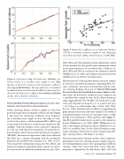
     Figure 4. Attenuation (top) and backscatter (bottom) coef- ficients (Coef.) of a cancellous bone sample in vitro (blue curves). A linear fit is shown for the attenuation coefficient data (top red dashed line). The slope of the line is normalized broadband ultrasound attenuation (nBUA). A power law fit is shown for the backscatter coefficient data (bottom red dashed curve). cmSr, centimeter steradians.
Devices for Bone Fracture Risk Assessment to provide a foun- dation for clinical quantitative ultrasound in bone.
Studies involving human calcaneus samples in vitro have provided insight into determinants of BUA and SOS. Figure 4, top, shows the attenuation coefficient versus frequency for a cancellous bone sample in vitro. The slope of a lin- ear fit to these data is called normalized BUA (nBUA) and is measured in decibels per centimeter per megahertz (dB/ cmMHz). When the thickness of the calcaneus is unknown, as is the case with clinical measurements, the parameter re- ported is BUA, which is measured in decibels per megahertz (dB/MHz). Mechanical compression studies have indicated that BUA has a strong relationship with the mechanical properties of cancellous bone (Langton et al., 1996). A theo- retical model for relationships among the SOS, nBUA, and dispersion (frequency-dependent phase velocity) in cancel- lous bone has been validated in human bone in vitro (Wear,
Figure 5. Backscatter coefficient versus trabecular thickness (Tb.Th) in 43 human calcaneus samples in vitro. Reprinted, from Wear and Laib, (2003), with permission, © 2003 IEEE.
2000). BUA and SOS primarily provide information related to bone quantity but also provide some information related to the microarchitecture of cancellous bone (Chaffai et al., 2002). BUA and SOS are sensitive to the volumetric density (Hoffmeister et al., 2000) and collagen and mineral content (Hoffmeister et al., 2002) of cancellous bone.
Measurements of scattering from human calcaneus samples in vitro have elucidated mechanisms underlying BUA (be- cause scattering is one source of attenuation) and the clini- cal scattering findings discussed in Clinical Ultrasound Devices for Bone Fracture Risk Assessment. Figure 4, bot- tom, shows the backscatter coefficient versus frequency for a cancellous bone sample in vitro. In the clinical frequency range (approximately 300-700 kHz), the backscatter coeffi- cient \\\\\\\[η(f)\\\\\\\] depends on frequency (f) as a power law \\\\\\\[η(f) = Af n\\\\\\\] when n is a little higher than 3 (Wear, 1999; Chaf- fai et al., 2000). The backscatter coefficient is approximately proportional to the mean trabecular thickness (the width of mineralized tubular structures in cancellous bone; see Fig- ure 1b) to the third power (Wear and Laib, 2003; Figure 5). Like BUA and SOS, backscatter is sensitive to the volumetric density (Hoffmeister et al., 2000), BMD (Hoffmeister et al., 2006), and collagen and mineral content (Hoffmeister et al., 2002) in cancellous bone. In the clinical frequency range, single scattering is believed to be much stronger than mul- tiple scattering (Wear, 1999; Haiat et al., 2008).
One interesting feature of porous media is that a single lon- gitudinal pressure wave entering a porous sample can gener- ate two longitudinal pressure waves propagating at different velocities (called “fast” waves and “slow” waves). This phe- nomenon is explained by the Biot theory (Biot, 1956) and
 Summer 2018 | Acoustics Today | 37
