
Page 41 - Summer 2018
P. 41
Over the past two decades, the determination of guided modes and identification of cortical bone waveguide characteristics has sparked increased investigation of signal-processing ap- proaches, modeling, and inverse problem solving. At least two cortical devices have appeared on the market and have been tested in clinical studies (Barkmann et al., 2000).
With earlier approaches, a one transmitter-one receiver con- figuration was implemented in which wave velocity could be computed from the separation distance and the measure- ment of the time of flight (TOF) between transmitted and re- ceived signals. The TOF technique has been used to evaluate the velocity of the first arrival signal (FAS), which is defined as the first component of the signal that emerges from noise. The FAS contains relevant information on the microstruc- tural and material properties (Talmant et al., 2011). How- ever, it cannot be easily predicted by analytical methods and therefore is difficult to infer from waveguide characteristics. FAS is a transient mode, consistent with a lateral longitudi- nal wave (which propagates at the longitudinal bulk veloc- ity) when the ratio of the cortical thickness to the acoustic wavelength is much greater than 1 or the S0 Lamb mode for a plate if the acoustic wavelength is not much greater than 1 (Talmant et al., 2011).
FAS velocity evaluation may be improved by making TOF measurements at multiple receiver positions, either by moving the receiver or by using a multielement transduc- er array. In addition to the FAS, a slower waveform has been isolated and interpreted as a fundamental flexural guided wave (FFGW; equivalent to A0 Lamb mode for a plate; Nicholson et al., 2002). The dispersion characteristics (frequency-related phase velocity variations) of this mode are very sensitive to cortical thickness (Moilanen et al., 2007).
One AT device uses two frequencies. Measurements at 100 kHz strongly depend on the cortical thickness, whereas measurements at 1 MHz depend mainly on propagation parameters of the bulk longitudinal wave (mass density and stiffness). Researchers claim that dual-frequency AT ultra- sound can detect early changes induced by osteoporosis more clearly than can single-frequency AT ultrasound (Sar- vazyan et al., 2009).
These analyses of AT signals were restricted to analyzing a single waveform, either the FAS or the FFGW. Howev- er, an infinite number of guided waves can exist. Multiple modes contain more information but are more difficult to interpret. Each mode interferes with every other mode, and
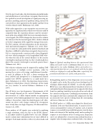
Figure 8. Optimal matching between the experimental data (dots) and Lamb modes (continuous lines) for an ex vivo (top) and in vivo radius (bottom). Inliers and outliers (i.e., experimental data that are not explained with the free-plate model) are displayed as red and blue dots, respectively. Re- printed from Bochud et al. (2016), with permission.
distinguishing modes or their dispersion curves in record- ed signals can be tricky. Researchers have developed an AT technique, bidirectional AT (BDAT), to solve this problem. BDAT uses a one-dimensional linear transducer array to re- cord guided modes that propagate in two opposite directions from two emitting transducer arrays placed on each side of the central receiving array. Combining measurements from two opposite directions automatically compensates for bias on measured wave speeds resulting from the surrounding soft tissues (Moreau et al., 2014).
The BDAT probe is a 1-MHz array adapted to clinical mea- surements at the one-third distal radius. It consists of 24 re- ceivers surrounded by 2 arrays of 5 emitters each. A matrix response is recorded by repeatedly firing pulses into the bone from each element of the emitting arrays and recording the response at each element of the receiving array. Dispersion curves are obtained by a reconstruction method based on a singular value decomposition combined with a two-dimen-
Summer 2018 | Acoustics Today | 39
