
Page 38 - Summer 2018
P. 38
Quantitative Ultrasound and Osteoporosis
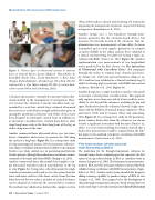
Figure 3. Various types of ultrasound systems to measure bone: a: General Electric Lunar (Madison, WI) Achilles®; b: BeamMed (Petah Tikva, Israel) MiniOmni®; c: Bone Index (Kuopio, Finland) Bindex®; d: CyberLogic (New York, NY) Ul- traScan 650®; e: Oyo (Kyoto, Japan) LD-100®; f: custom back- scatter system (Wear and Armstrong, 2001).
Calcaneal ultrasound is currently the most prevalent ultra- sound method in the management of osteoporosis. How- ever, because the calcaneus is mostly cancellous bone sur- rounded by a very thin cortical layer, calcaneal ultrasound does not assess cortical bone strength and therefore gives an incomplete prediction of fracture risk. Other devices have been designed to interrogate cortical bone in addition to, or instead of, cancellous bone. Cortical bone devices often target long bones such as the tibia (long bone of the leg) or radius (long bone of the arm).
Another commercial bone ultrasound device uses two trans- ducers (transmitter and receiver) to measure the SOS along the cortex of a long bone (Figure 3b). In a retrospective study of 254 postmenopausal women, SOS measurements at the fore- arm, finger, or midfoot demonstrated an ability to discriminate fracture cases from control cases in postmenopausal forearm fracture patients, although the performance was inferior to mea- surements of the spine and femur BMDs (Knapp et al., 2002). Another commercial bone ultrasound device applies a sin- gle ultrasound transducer placed perpendicular to the leg or arm (Figure 3c). Using pulse-echo ultrasound, the single transducer transmits a pulse and receives the echoes from the outer and inner surfaces of the bone cortex. From the time delay between the two echoes, an index of cortical thickness can be obtained if a value for the cortical SOS is assumed. The method was validated on human tibia samples in vitro
(Wear, 2003) and in a clinical trial involving 572 women for improving the management of patients suspected of having osteoporosis (Karjalainen et al., 2016).
Another design uses a two-transducer through-trans- mission geometry, like the calcaneus-based devices, but measures the forearm instead of the calcaneus. One im- plementation uses measurements of time-delay (between transmitted and received signals) parameters to estimate an index of BMD in the radius and has been validated in a clinical trial involving 60 adults for having a high cor- relation with BMD (Stein et al., 2013; Figure 3d). Another implementation uses measurements of two longitudinal waves predicted by the Biot theory (see The Interaction of Ultrasound with Cancellous Bone) that propagate through the radius to estimate bone density and elastic- ity (Otani et al., 2009) and cortical thickness (Mano et al., 2015) and has been validated in a clinical trial involving 93 adults for having high correlations with BMD and cortical thickness (Breban et al., 2010; Figure 3e).
Another design uses a single transducer in pulse-echo mode to measure scattering from cancellous bone. This approach only requires access from one side of a bone and has acces- sibility to sites beyond the calcaneus, including the hip and spine. Backscatter from the calcaneus showed a high corre- lation with the BMD in 10 normal human volunteers (Wear and Garra, 1998) and 47 women (Wear and Armstrong, 2001; Figure 3f). In a retrospective study of 210 postmeno- pausal women, backscatter from the calcaneus was shown to have a significant association with fractures (Roux et al., 2001). A recent study involving 342 women showed that a backscatter measurement could be acquired from the lum- bar spine in vivo and had a moderate correlation with BMD measurement (Conversano et al., 2015)
The Interaction of Ultrasound
with Cancellous Bone
The motivation for the through-transmission calcaneus ul- trasound comes from a seminal paper that represents the first report of an age-related decline in BUA in cancellous bone in women (Langton et al., 1984). The first paper to report measure- ments of BUA and SOS in human volunteers showed that both exhibited strong correlations with BMD measurements (Zagze- bski et al., 1991). Another early system extended this design by adding scanning capability to produce images of BUA in vivo (Laugier et al., 1996). These papers led to the development of clinical through-transmission calcaneus-based systems that were used in early large-scale trials mentioned in Clinical Ultrasound
36 | Acoustics Today | Summer 2018
