
Page 26 - Spring2019
P. 26
Theracoustic Cavitation
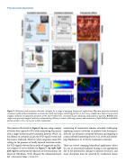
Figure 5. Nucleation and cavitation detection strategies for a range of emerging theracoustic applications. Top row: passively monitored cavitation-mediated thermal ablation nucleated by locally injected gas-stabilizing particles in liver tissue. Center row: dual-array passively mapped cavitation-mediated fractionation of the intervertebral disc, nucleated by gas-stabilizing solid polymeric particles. Bottom row: single-array passively mapped cavitation-enhanced drug delivery to tumors, following systemic administration of lipid-shelled microbubbles and an oncolytic virus. See text for a fuller explanation.
This process is illustrated in Figure 5, top row, using a sample of bovine liver exposed to FUS while monitoring the process with a single-element passive-cavitation detector (PCD). In the absence of cavitation nuclei, the PCD signal is weak and the tissue is unaltered. However, when using these same FUS settings in the presence of locally injected cavitation nuclei, the PCD signal is elevated by an order of magnitude and dis- tinct regions of tissue ablation (in Figure 5, top right, light pink regions correspond to exposures at three locations) are observed (Hockham, 2013). Because the ultrasound-based 24 | Acoustics Today | Spring 2019
monitoring of temperature remains extremely challenging, exploiting acoustic cavitation to mediate tissue heating en- ables the use of passive cavitation detection and mapping as a means of both monitoring (Jensen et al., 2013) and control- ling (Hockham et al., 2010) the treatment in real time.
There are several emerging biomedical applications where the use of ultrasound-mediated heating is not appropriate due to the potential for damage to adjacent structures, and tissue disruption must be achieved by mechanical means
