
Page 35 - Winter2021
P. 35
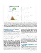
  Figure 7. Top left: Gaussian distributions of native frequency and bandwidth representing 13 fibrotic patients. Color scale, probability density (unitless), Top right: two-dimensional (2-D) plot representing 26 patients with respect to the mean bandwidth and native frequency of their vertical artifacts, Bottom left: histograms of the vertical artifact total intensity as obtained from fibrotic (red) and nonfibrotic (light green) patients. Itot, total intensity Bottom right: 2-D plot representing 26 patients with respect to the mean bandwidth and native frequency of their vertical artifacts after a threshold of vertical artifact intensity was applied.
Multiple Ultrasound-Based Parameters to Increase Specificity
Another highly relevant way to achieve specificity is to combine multiple parameters. In that sense, the physics- based ultrasound parameters described in Parameters Related to Scattering and Diffusion, which are related to diffusion, scattering models, and attenuation, respec- tively, are not redundant and will reflect different and independent properties of the lung structure. Using them in combination is a path that should be considered to increase the specificity of diagnosis.
For example, pulmonary fibrosis and pulmonary edema both increase the diffusion constant because they both lower the alveolar density. As a result, the measurement of the SMFP is not sufficient to discriminate between them (Figure 6). The BFS, on the other hand, is lower in edema- tous lungs (low attenuation) than in fibrotic lungs (high attenuation). Moreover, vertical artifact native frequency and bandwidth provide an indirect estimation of the acous- tic channel size and geometries, whereas the total intensity helps in discriminating the channel content (Figure 7). As
a consequence of these differences in approaches, it is pos- sible to envision a multiparameter space that would allow simultaneous quantifying of alveolar density, mean alveo- lar size, and tissue characteristics (interstitial fluid, fibrotic tissue, or air).
This new research suggests that ultrasound can be uti- lized to monitor and diagnose lung diseases. Clinical applications are currently limited to semiquantitative methods that can, however, provide important informa- tion on the level of aeration of the superficial lung. But promising developments (Demi, 2020; Mento et al., 2020; Mohanty et al., 2020; Lye et al., 2021) suggest that quan- titative and specific ultrasound characterization of lung tissue is within reach.
The Potential of Portability: What Could It Look Like?
Compared with other diagnostic modalities, ultrasound has the significant advantage of allowing relatively inexpensive and portable care (Everbach, 2006; Ketterling and Silver- man, 2017; Simon et al., 2017; Smallwood and Dachsel,
Winter 2021 • Acoustics Today 35
