
Page 33 - Winter2021
P. 33
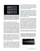
  Figure 4. Examples of lung ultrasound images obtained with a multifrequency imaging approach in three different patients. From left to right, images formed with ultrasound pulses at a different center frequency (i.e., 3, 4, 5, and 6 MHz). Light blue arrows, pleural line; red arrows, vertical artifacts.
Scattering and Diffusion by the Alveoli
In the lungs, alveoli scatter ultrasound waves due to their shape, size, and the strong acoustic impedance difference between air and tissue. The portion of the wave that is able to penetrate the lungs is scattered back in the form of a highly complex signal. This is a significant challenge because it prevents ultrasound imaging. However, each scat- tering event is, in fact, an opportunity, leaving the signature of the microstructure within the ultrasound signals. One way to leverage this information is to use a mathematical analysis to extract information from raw ultrasound data as opposed to interpreting features from ultrasound images in which the ultrasound data have already been processed.
Because of the large numbers of scattering events by the millions of air-filled alveoli, ultrasound waves do not propagate straight at a constant speed. Instead, they are subjected to diffusion. One can exploit the physics of diffusion and scattering to extract new parameters that could be used as quantitative biomarkers for lung dis- eases such as fibrosis or edema.
In a process dominated by multiple scattering and dif- fusion, the incoherent contribution of the ultrasound signals is stronger. To describe the principle of how this could be leveraged, one can use an analogy with a group a people walking in a forest. The walkers walk straight from tree to tree but randomly change direction at each tree encounter. In a very dense forest (e.g., healthy lung),
the walkers would quickly forget where they came from after a few tree encounters. As a group, they would make slow progress in the forest. In contrast, in a sparse forest (diseased lung), they would make quicker progress and the group would quickly spread apart.
Parameters Related to Scattering and Diffusion
In the lungs, the air-filled alveoli play the role of the aforementioned trees. Measuring the rate of spread of the diffusive halo (Figure 5) provides an indirect mea- surement of the density of alveoli, which is affected by interstitial lung diseases such as pulmonary fibrosis and pulmonary edema. The rate of growth of the diffusive halo is the diffusion constant.
For spherical air-filled scatterers, the diffusion constant is proportional to the scattering mean free path (SMFP), which represents the mean distance between scattering events and is a measure of the mean distance between healthy alveoli (Mohanty et al., 2017). In a rodent study on rats with pulmonary fibrosis, rats with pulmonary edema, and healthy rats, it was demonstrated that the SMFP was significantly lower in healthy animals than in animals with fibrosis or edema (Figure 6) (Mohanty et al., 2020). It has also been showed that the SMFP was signifi- cantly correlated to the severity of fibrosis as measured by histology, the gold standard for pulmonary fibrosis.
In addition to the diffusion constant and SMFP, the ultra- sound backscatter coefficient (BSC) and ultrasound signal
 Figure 5. Growth of the diffusive halo over time in a healthy lung (left) and in a lung with edema (right). Because the density of air-filled alveoli is lower in edematous lungs, the diffusive halo grows faster. Color scale, normalized incoherent intensity.
 Winter 2021 • Acoustics Today 33
