
Page 31 - Winter2021
P. 31
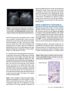
  Figure 2. Lung ultrasound images from two different Covid-19 patients displaying consolidations and extended vertical artifacts. The white dashed lines overlaid on each image are indicative of the original location of the pleural line.
which ultrasound waves can penetrate and in which they are trapped. The pinball effect in that case occurs inside these structures because the ultrasound wave finds itself surrounded by “air walls.” Given the size of these structures, which can be close to the wavelength at the imaging frequency, resonance rather than reverberation phenomena can occur. This currently represents the main hypothesis behind the visualization of vertical artifacts (Demi et al., 2020; Mento et al., 2020).
It is clinically relevant that although horizontal artifacts are generally associated with a healthy lung, a correlation exists between vertical artifacts and many lung condi- tions, including Covid-19 (Figure 2) (Soldati et al., 2020). It should also be noted that lung ultrasound imaging can only detect alterations that occur close the lung surface. That is the only part of the lung that can be explored because the presence of air significantly hinders ultra- sound propagation. In the case of a more critical lack of aeration, such as with lung consolidation, ultrasound waves can penetrate deeper into the lung and the sur- face of discontinuity between dense nonaerated areas and aerated areas moves deeper with respect to the original location of the pleural line.
Figure 2 shows examples of lung ultrasound images displaying consolidations in a Covid-19 patient. Consoli- dations can be defined as large areas (at least one order of magnitude larger than the wavelength at the imag- ing frequency) of the lung, originally occupied by air,
that become filled with denser media such as blood and inflammatory liquids. These images show that beyond consolidations, vertical artifacts extend from the bottom edge of the hypoechoic areas (displaying low intensi- ties). These are signs of partial, although pathological, aeration levels. Consolidations are typical in Covid-19 patients but do not represent a specific sign (Soldati et al., 2020) because they can be observed with many other lung conditions.
Chronic Lung Diseases: The Example of Pulmonary Fibrosis and Pulmonary Edema Idiopathic pulmonary fibrosis (IPF) is a chronic condition that affects 200,000 Americans (Raghu et al., 2018). With IPF, scarring in lung tissue occurs progressively (Figure 3), leading to serious breathing difficulties. IPF is lethal but can be mitigated with new drugs. Pulmonary fibrosis can be monitored by periodic high-resolution computed tomography (HRCT) scanning and pulmonary function tests. HRCT is costly and ionizing and pulmonary func- tion tests need to be performed in a hospital for accuracy.
Developing quantitative ultrasound methods for the quantification of pulmonary fibrosis would enable an inexpensive point-of-care monitoring of the disease pro- gression and response to treatment. Pulmonary edema is another interstitial lung disease. It is related to heart
 Figure 3. Top: histology of a healthy rat lung. The alveolar density is high. Bottom: histology of a fibrotic rat lung. The alveolar density is significantly lower.
 Winter 2021 • Acoustics Today 31
