
Page 32 - Winter2021
P. 32
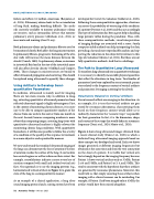
NOVEL METHODS IN LUNG ULTRASOUND
failure and affects 6.2 million Americans (Benjamin et al., 2019). Pulmonary edema leads to the accumulation of lung fluid, making breathing difficult. The meth- ods currently available to monitor pulmonary edema are invasive, such as intracardiac devices that monitor pulmonary artery pressure (Abraham et al., 2011) or inaccurate and ionizing (chest X-ray).
Both pulmonary edema and pulmonary fibrosis need to be monitored closely. Both affect the lung microstructure. In pulmonary fibrosis, progressive thickening of the alve- olar walls will increase the mean distance between the alveoli (Crouch, 1990). In pulmonary edema, an increase in interstitial fluid and in the size of the interstitial space will also affect alveolar density (Gehlbach and Geppert, 2004). Those changes in microstructure are bound to affect ultrasound propagation and scattering. This should be explored using ultrasound to quantify those changes.
Using Artifacts to Develop Semi- quantitative Parameters
In medicine, ultrasound is mainly used for imaging. There are two main reasons why, in addition to imag- ing, extracting quantitative ultrasound parameters from reflected ultrasound signals is highly advantageous. First, in the context of monitoring chronic diseases, it is neces- sary to be able to compare quantitative markers of the disease from one week to the next or from one month to the next. Second, because comparing numbers is more robust than comparing images, assessing lung tissue with quantitative ultrasound markers is highly relevant for monitoring chronic lung conditions. With quantitative biomarkers, it will become possible to follow the severity of a condition or the quality of the response to treatment in a more objective and reproducible manner.
We now understand that standard ultrasound imaging of the lungs can demonstrate the loss of aeration if the loss of aeration reaches the surface of the lung. It can localize less aerated regions as well as inform on their extent. For example, consolidations indicate a more severe loss of aeration compared with small and isolated vertical arti- facts. One approach is to use the imaging patterns (e.g., horizontal and vertical artifacts in Figure 1) to grade the state of the lung in a semiquantitative manner.
As an example of a clinical application, a lung ultra- sound imaging protocol and a scoring system have been
developed for Covid-19 evaluation (Soldati et al., 2020). Following these semiquantitative approaches, clinicians can assess the probability of worsening of the condition of a patient (Perrone et al., 2020) and thus act accordingly. This type of information has been a great help in handling large patients’ influx during the pandemic. Thus, with these semiquantitative methods, visual interpretation of imaging findings are associated with a score. Surely, computer-aided methods can help interpreting the data, providing a fast and more reproducible analysis and sup- porting the reduction in the subjectivity intrinsic to this type of evaluation (Mento et al., 2021). But these are not fully quantitative methods. And that is a challenge.
The Path to Quantitative Lung Ultrasound
To transform lung ultrasound into a quantitative technique, it is necessary to identify measurable physical quantities that reflect the alterations in lung tissue. Two families of parameters have been explored by recent research: param- eters related to the frequency content of vertical artifacts and parameters leveraging scattering by the alveoli.
Parameters Related to Vertical Artifacts
Recent research efforts are emerging in this direction. As an example, if it is true that vertical artifacts are gen- erated by resonance phenomena, characterizing them based on their frequency content would allow us to indirectly characterize the “acoustic traps” responsible for their generation. In fact, it is the dimension, shape, and content of these traps that would define its resonance frequency (Demi et al., 2020; Mento et al., 2020).
Figure 4 shows lung ultrasound images obtained from a recent clinical study (Demi et al., 2020) in which a dedicated lung ultrasound imaging approach based on these concepts was developed and tested. Each row shows images generated at different imaging frequencies but obtained at the same time and from the very same point on the chest of a patient. It is visible that vertical arti- facts strongly depend on the imaging frequency because Patient 1 shows vertical artifacts only at 3 MHz, Patient 2 at 4 and 5 MHz, and Patient 3 at 3, 4, and 5 MHz. This result is consistent with the hypothesis that these artifacts are generated by resonance phenomena. Moreover, this result tells us that simply counting these artifacts when imaging with a clinical scanner can be misleading. For example, if Patient 1 had been imaged only at 4 MHz, the artifact would have been missed altogether.
32 Acoustics Today • Winter 2021
