
Page 30 - Winter2021
P. 30
NOVEL METHODS IN LUNG ULTRASOUND
the probe) and one acoustic interface and not by the interac- tion between “echoes” and an acoustic interface.
These two hypotheses are valid for most biological tissues. As an example, in tissues such as blood, brain, muscle, fat, breast, heart, kidney, liver, and spleen, errors smaller than ±7.5% would be made assuming a homogeneous speed of 1,546m/s (Szabo, 2004). This also implies that echo intensities are generally small, given that the reflection coefficients at these interfaces are small (Szabo, 2004).
In those tissues, higher order scattering can be ignored.
With higher order scattering, the linear relationship between time of flight (i.e., the interval between the transmission of the ultrasound pulse and the reception of its echo) and the actual distance of the interface responsible for that echo is lost. In other words, strong higher order scattering means that the echolocation principle is fundamentally inappli- cable. This is precisely what happens in the lungs.
Clinical Lung Ultrasound
The fact that echolocation cannot be used in lungs is pre- sumably the reason why the interest from the scientific community in lung ultrasound has faded until new devel- opments from the clinical world came in the late 1990s (Lichtenstein et al., 1997). Fortunately, blind to the fact that the basic assumptions behind standard ultrasound imag- ing did not apply to lung tissue, clinicians began imaging lungs with ultrasound clinical scanners and started to report and describe the presence of “signs” that appeared on their screens with a wide variety of lung diseases.
All lung images exhibited imaging artifacts, but striking differences were observed between ultrasound images in healthy aerated lungs and in diseased lungs (Soldati et al., 2016). An imaging artifact is a feature that appears in an image (intended as an anatomical representation of internal body parts) but that is not actually present in the imaged volume. It is, in fact, the imaging system that generates these artifacts because it applies predefined processing operations to signals that do not verify the assumptions made by the system.
Although conventional ultrasound images fail to pro- vide an anatomical description of the lungs, artifacts still convey information in the sense that they signal that something particular is happening. The question is, what use can be made of these artifacts?
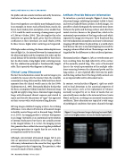
Artifacts Provide Relevant Information
To introduce a practical example, Figure 1 shows lung ultrasound images exhibiting horizontal (called A-lines) and vertical (called B-lines) artifacts. In both images, it is possible to clearly distinguish the intercostal tissue layers (between the probe and the lung surface) and a first hori- zontal structure, known as the pleural line, which is the anatomical representation of the lung surface and which separates the image into two parts. Up to the pleural line, the two fundamental assumptions necessary to ultrasound imaging still hold. Below the pleural line is a world of arti- facts because the wave is encountering lung tissue and the imaging volume is filled with air. These images can be dis- tinguished by the differences in their artifactual patterns.
Horizontal artifacts (Figure 1, left) are reverberation arti- facts resulting from the high reflectivity of the surface of the normally aerated lung. This series of horizontal lines is the visual representation of the multiple reflec- tions occurring between the ultrasound probe and the lung surface. A sort of pinball effect between the probe and the lung surface that, if the lung is fully aerated, acts as an impenetrable wall to ultrasound waves.
In contrast, vertical artifacts (Figure 1, right) are attrib- uted to local alterations in the acoustical properties of the lung surface, such as the replacement of volumes normally occupied by air in favor of media that are acoustically more similar to the intercostal tissue (water, blood, and tissue), resulting in an increased transmission coefficient. These alterations are typical of a wide range of pathological conditions that opens channels through
Figure 1. Lung ultrasound images displaying horizontal (left) and vertical (right) artifacts. See text for detailed explanations.
30 Acoustics Today • Winter 2021
