
Page 51 - 2017Spring
P. 51
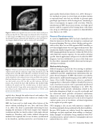
 Figure 4. Midsection sagittal view of an in utero mouse embryo on embryonic day (E) 12.5. The image was obtained with a Visual Son- ics 3100 using a 40-MHz linear array transducer. It was acquired at the Skirball Institute of Biomolecular Medicine at New York Univer- sity Langone Medical Center and is courtesy of O. Aristizábal.
quire another batch of data (Chérin et al., 2006). Retrospec- tive methods are prone to errors from out-of-plane motion or unsteady heart rates but are still able to generate quite good high-speed movies of the beating heart, considering it takes several minutes to acquire a full set of data. With the introduction of HFU linear-array systems, true high-speed movies could be acquired of the beating heart, with frame rates of up to 1,000 frames per second over a limited field of view (Foster et al., 2009).
Recent Developments
As noted in Applications, HFU has had a foothold in the clinic for many years but has not emerged as a common tool for general imaging. This was a classic chicken-and-egg situ- ation in that there was no FDA-approved HFU machine so new clinical applications were not aggressively pursued. This situation has finally changed because VisualSonics released a HFU imaging platform that operates within the FDA 510(k) safety limits for acoustic exposure. It is too early to tell what specific clinical applications will prove to be ideal for HFU diagnosis, but they will likely be in areas of the body near the skin where commonly available ultrasound systems pro- vided poor resolution.
Multimodality Imaging
Medical imaging in general has been moving to multimo- dality imaging approaches where two imaging methods are combined to provide complementary information that im- proves disease diagnosis. In HFU, this trend is seen with the photoacoustic, small-animal imaging system sold by Visual- Sonics (Vevo LAZR). Photoacoustics refers to a method of imaging where a short pulse of light is absorbed by tissue in such a way that an acoustic wave is generated and detected by an ultrasound transducer (Emelianov et al., 2009). The photoacoustic mode provides information related to the molecular absorption of light at a specific wavelength and the ultrasound mode provides mechanical information about tissue structure. The image information from each modality is coregistered and can be used to detect targeted contrast agents, areas of vasculature, or blood oxygenation in the regions of specific organs.
IVUS imaging is also an area with numerous examples of the ultrasound probe being combined with other modalities. Because IVUS probes are catheter based, the engineering challenges are quite significant when adding a second mo- dality. Examples of modalities being combined with IVUS are photoacoustic, optical coherence tomography and near infrared (Ma et al., 2016).
 Figure 5. Volume reconstructions (a-e) and the corresponding mid- sagittal sections (f-j) for embryonic stages E10.5 (a and f), E11.5 (b and g), E12.5 (c and h), E13.5 (d and i), and E14.5 (e and j) using data obtained with a 40-MHz annular array. The brain ventricles are easily identified in the embryo because they are filled with anechoic fluid and therefore have a high contrast relative to surrounding tis- sue. BA, basilar artery; EY, eye; ER, ear; FB, forebrain; FF, facial features; FL, forelimb; H, heart; HB, hindbrain; HL, hindlimb; MB, midbrain; SC, spinal cord; VA, vertebral artery; 3v, third ventricle; 4v, fourth ventricle. Large white arrows (g and j) indicate interso- mitic blood vessels. Reprinted from Aristizábal et al. (2013), with permission from Elsevier.
sagittal slices through the midsection of each embryo. The growth and development of the embryo are clearly visual- ized as the embryo ages.
HFU has been used to study many other features of the mouse embryo including the eyes, liver, and heart (Zhou et al., 2002). The heart is particularly interesting because imaging the mechanics of the heart requires fine temporal resolution and this was hard to achieve with single-element transducers. Therefore, early studies utilized retrospective methods that acquired data in one location, synchronized to the heartbeat, and then moved to a new location to ac-
 Spring 2017 | Acoustics Today | 49
