
Page 52 - 2017Spring
P. 52
High-Frequency Ultrasound Applications
High-Speed Imaging
Although traditional Doppler is used extensively for blood flow measurements in general, low-megahertz ultrasound imaging, it is not commonly used to image the eye. The primary reason for this is that traditional Doppler modes generally exceed the stringent FDA 510(k) ophthalmic ul- trasound exposure limits. Thus, very little is known about real-time blood flow in the eye as it relates to healthy and diseased eyes. HFU linear arrays and an emerging technique known as plane-wave imaging may offer a means to over- come the above obstacles and provide new blood flow-relat- ed information for studying and diagnosing ophthalmic dis- eases such as glaucoma, macular degeneration, retinopathy of prematurity, and tumors. The technique can also be used to image cardiovascular function in mice or other small ani- mals.
High-speed plane-wave or diverging-wave imaging has been enabled by advances in ultrasound equipment (Tanter and Fink, 2014). In a conventionally operated linear array, groups of adjacent elements emit a focused, converging wave front, and the elements must wait until echoes are received before the next group can be excited. This process is then iterated across the length of the array to acquire one image. In plane- wave imaging, all elements of the array fire to emit a single unfocused wave front. Plane-wave imaging thus permits full-frame transmission rates up to the round-trip propa- gation time of a transmit-and-receive event. For instance, a 2-cm depth from the face of a transducer could be imaged at 36,000 frames per second. High-speed plane-wave imag- ing is well suited to cases with transient high-speed motion or blood flow such as cardiovascular imaging or shear-wave imaging (Tanter and Fink, 2014).
The trade-off with plane-wave imaging is that because the transmit beam is unfocused, the resolution is degraded and the acoustic intensity is reduced. This can be partially over- come by transmitting a batch of electronically steered plane waves and then summing (i.e., compounding) the resulting beamformed data. As the number of transmission angles per image increases, the SNR and resolution improve, al- though the overall effective frame rate necessarily decreases. For instance, an experiment may entail emitting batches of 10 plane waves at a rate of 20 kHz over a ±15° angle range. Because 10 angles are compounded to form one image, the final effective frame rate is 2 kHz.
Plane-wave imaging permits near-instantaneous Doppler flow information to be obtained throughout a full-image
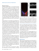
Figure 6. Image of blood flow in the region of the optic nerve in a normal human eye. The image was obtained using a technique called coherent compound plane-wave imaging (Urs et al., 2016). In this case, 20,000 images per second were acquired at 5 angles using an 18-MHz linear array. Red, arterial flow; blue, venous flow. The Dop- pler spectrograms on the right display flow velocities in the central reti- nal vein (1) and posterior ciliary artery (2) over a 1.6-second period.
frame. For a similar number of transmissions used in plane- wave imaging, traditional Doppler methods are only able to obtain flow information in a small region because the transmit beam is focused using a subset of array elements. Therefore, plane-wave imaging is able to provide detailed flow information but at a lower acoustic intensity than with traditional Doppler approaches.
An example of the plane-wave technique applied to the eye using an 18-MHz linear array is shown in Figure 6. It shows blood flow in the major retrobulbar vessels (central retinal artery and vein, ophthalmic artery, short and long ciliary ar- teries, and vortex vein) as well as flow in the choroid. Most importantly, the instantaneous and temporally averaged acoustic intensities are well within FDA 510(k) ophthalmic safety limits (Urs et al., 2016).
Conclusions
HFU represents the gradual progression of ultrasound tech- nology to higher and higher frequencies. The technology in use for HFU has tended to lag behind what is used on a daily basis for low-megahertz clinical ultrasound. The early HFU systems were focused on ophthalmic, small-animal, and intravascular applications, and the devices filled a special- ized niche. Today, we are finally seeing HFU enter the main- stream of clinical ultrasound with FDA approval of a HFU linear-array system for clinical use. At the same time, im-
50 | Acoustics Today | Spring 2017
