
Page 18 - Jan2013
P. 18
bubble oscillations and vessel displacements were observed on microsecond time scales.
For these experiments, approved by the University of Washington institutional animal care and use committee (IACUC), a rat mesentery was selected as the animal tissue model because it contains thin and transparent regions, allowing easy observations of its microvasculature under light microscopy. Experiments were conducted on vessels ranging from about 10-100 μm in diameter. These included arterioles, venules and capillaries. The confinement imposed by these vessels and surrounding tissue did not prevent bub- bles from undergoing large volumetric oscillations that included inertial collapses. In turn, vessels deformed on the same microsecond time scale as bubble oscillations. A typical observation of bubble dynamics is illustrated in Fig. 5. The tensile portion of the sound wave leads to microbubble growth, causing distention of the nearby vessel wall. The sub- sequent invagination of the vessel wall follows inertial bubble collapse. In this and most other cases, invagination appears more localized, suggesting higher stresses and strains than those resulting from distention. We could hypothesize, based on these findings, that invagination may be a principal mech- anism for bioeffects, at least in these types of vessels.34 Other vessels such as arteries are much stiffer than these vessels, and may not respond in the same manner. Still, most of the available evidence from in vivo studies indicates that vessel permeabilization effects occur principally in the microcircu- lation—that is, arterioles, venules and capillaries.
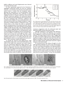
To quantify the observed vessel displacements, radial displacements of the point on the vessel wall closest to the center of the bubble were measured, and results obtained from four representative high-speed image sequences34 are plotted in Fig. 6. In each of these cases, distention was small relative to invagination. Moreover, vessel walls behaved sim- ilarly in that the average inward wall speed was around 9 m/s over the time range from 1.5–2.5 μs. Achieving such a veloc- ity over such a short time scale implies that this response was forced rather than evoked. In addition, vessels achieved their
maximum invaginations after the ultrasound pulse had passed and bubble motions had mostly ceased.
Although invagination is often greater than distension, we have also seen evidence that distention can be greater than invagination in small microvessels with high insonation pressures. The image sequence shown in Fig. 7 suggests that for small microvessels at high pressures, distention might be significant enough to rupture the vessel wall. However, this appears to be limited to small vessels under high pressures, in which the bubble expands significantly beyond the original diameters of the blood vessels.
We mentioned above that jetting is thought to be the other principal mechanism for some of the observed bioef- fects. We too observed clear evidence of jetting microbubbles in these vessels. Figure 8 illustrates one such observation. However, the jet was directed away from the nearby vessel wall, not towards it. In fact, in all of our data sets (over 20) in which jets can be clearly resolved, all of them were directed away from the nearest boundary.35 In Fig. 8 the microbubble
Fig. 7. A high-speed image sequence that shows expansion, collapse and re-expansion of a bubble cluster, accompanied by the observation of vessel distention and invagina- tion. Vessel diameter =17 m; ultrasound peak negative pressure = 7 MPa. The bubble fragments outside the vessel at 1.2 ms indicate the vessel was ruptured.
Fig. 8. Microjet formed in a 48 μm diameter microvessel under a peak negative pressure of 3 MPa.35 The scale bar represents 10 mm.
Fig. 6. Measurements of radial displacements of the vessel wall at the point closest to the bubble for four different experiments. Each marker denotes a measurement from a single image frame. Deflections toward the lumen were defined to be nega- tive. For each of these sequences, vessel invagination exceeded distention by a sig- nificant margin. The observed invaginations occurred after bubbles collapsed (at about 2 μs in the plot) and persisted even after bubbles rebounded.34
Microbubbles as Ultrasound Contrast Agents 17
